
Monoclonal gammopathy of unknown significance in kidney transplanted patients
Home | NDT Kidney Art Gallery
NDT Kidney Art Gallery
Welcome to NDT's Kidney Art Gallery
Welcome to NDT’s Kidney Art Gallery, a captivating visual journey into the intricate world of the kidneys!
This collection of stunning images submitted by NDT’s most creative readers and artists offers a unique glimpse into the complex structures and functions of the kidneys, showing the remarkable interplay of form and function in these vital organs. We invite you to explore the fascinating realm of the kidneys and see kidney health and disease from a new perspective.
Selected images will appear on the covers of NDT:
Display all Advanced microscopy Cells Digital art Drawing Electron microscopy Histology Imaging Urine & urine microscopy
12/54 elements


Author: Margaux Van Wynsberghe, France
Advanced microscopy
Info:
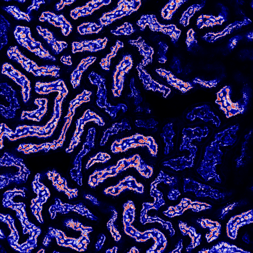
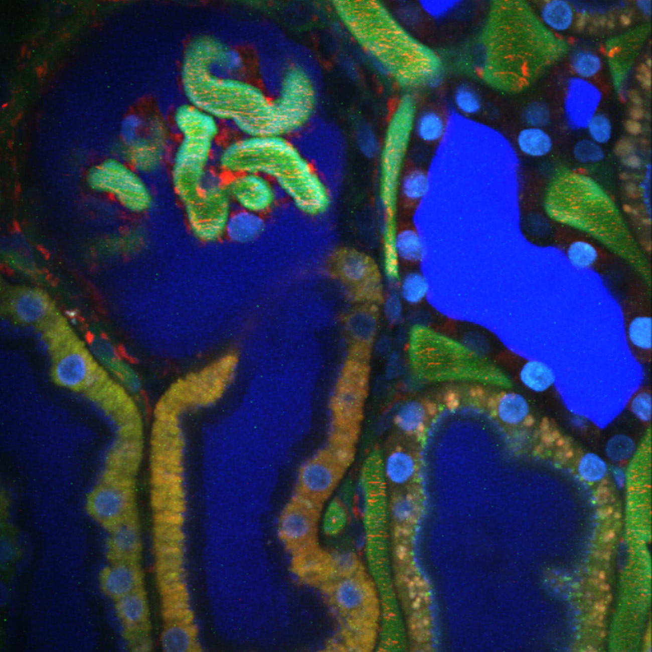
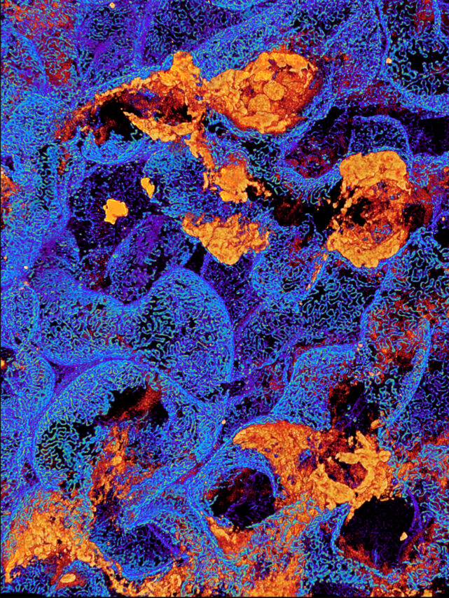
Glomerulus lover – Representative image showing immunofluorescent staining for nephrin (green), FAK (red) and DAPI (blue) on a piece of human nephrectomy.
From:
Margaux Van Wynsberghe, France
IMRB Inserm U955, Hôpital Henri Mondor

Author: Panagiotis Pateinakis, Greece
Histology
Info:
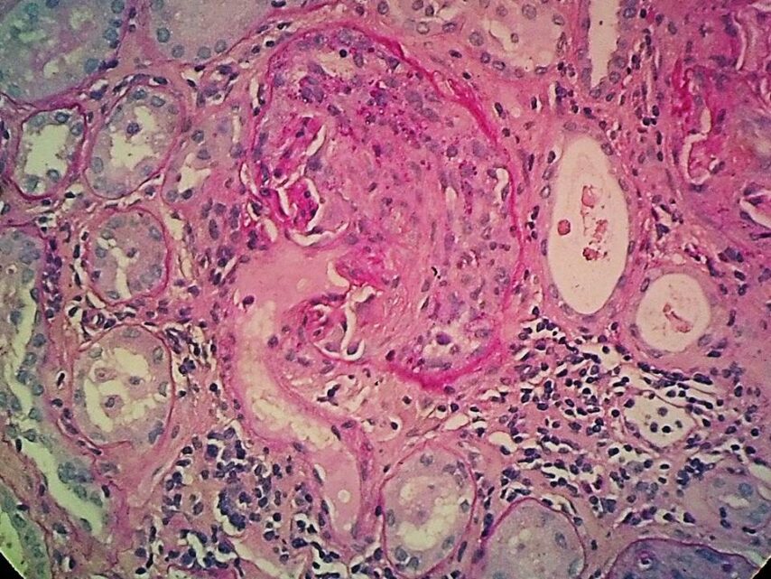
“Inception”- like, kidney-shaped nephron with a cellular crescent, from a patient with pauci-immune ANCA-associated renal vasculitis
From:
Panagiotis Pateinakis, Greece
Nephrology Department, “Papagergiou” General Hospital

Author: Maria Lambropoulou, Greece
Histology
Info:
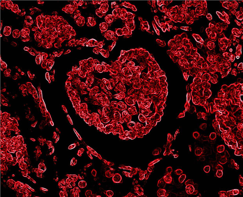
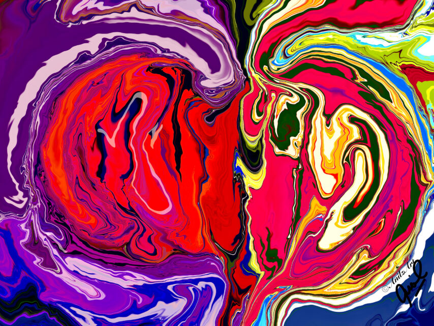
Kidney histological section in which a glomerulus forming the most iconic symbol of love; a big heart has been discovered in a hematoxylin-eosin slide (negative colors) after a hard day working on microscope
From:
Maria Lambropoulou, Greece
Pathologist & Professor of Histology-Embryology
Medical Department, Democritus University of Thrace, Alexandroupolis

Author: Maria Lambropoulou, Greece
Histology
Info:
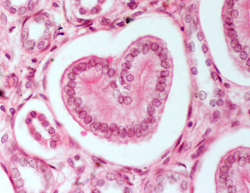
Kidney histological section in which the “heart” represents collecting tubule (hematoxylin-eosin staining, 200×)
From:
Maria Lambropoulou, Greece
Pathologist & Professor of Histology-Embryology
Medical Department, Democritus University of Thrace, Alexandroupolis

Author: Mayleen Laico, Philippines
Digital art
Info:
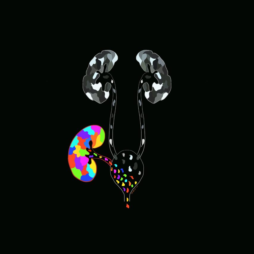
The Gift of Organ Donation
From:
Mayleen Laico, Philippines

Author: Oliver Kretz, Germany
Electron microscopy
Info:
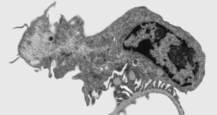
Transmission electron microscopy image of the murine renal filtration barrier. Random sections of the podocytes are sometimes reminiscent of prehistoric creatures.
From:
Oliver Kretz, Germany

Author: Yaoxian Xu | Rafael Kramann, Germany
Cells
Info:
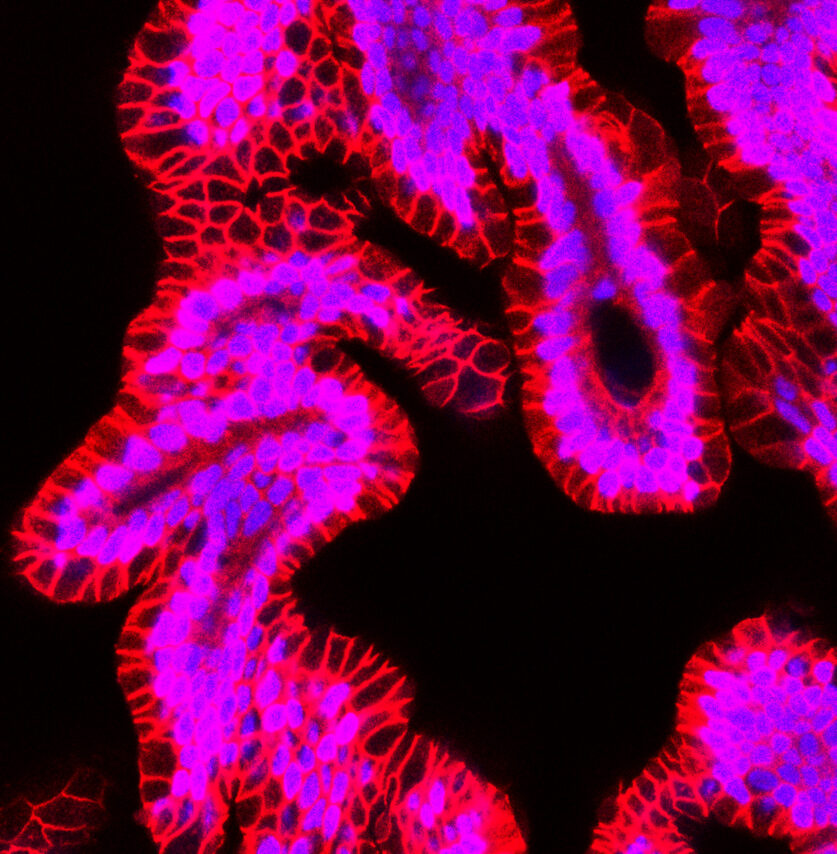
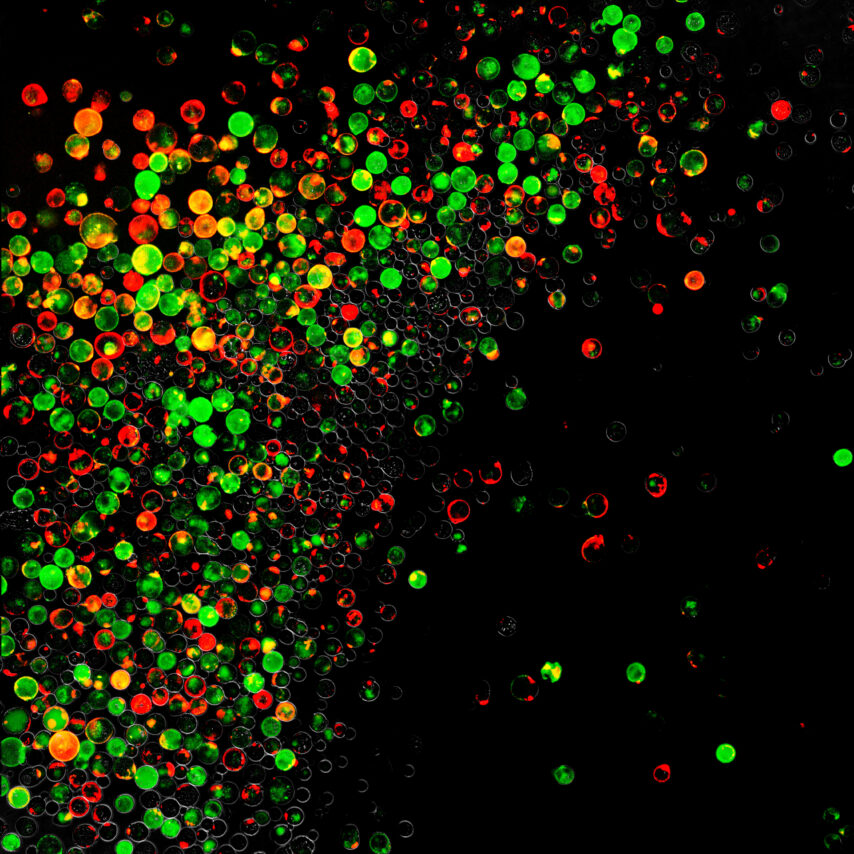
Adult human kidney organoids or tubuloids derived from CD24+ stem/progenitor cells. Red color in confocal image indicates E-cadherin (CDH1) staining, blue is DAPI.
From:
Yaoxian Xu and Rafael Kramann, Germany
Institute of Experimental Medicine and Systems Biology, Medical Clinic 2, Faculty of Medicine, RWTH Aachen University.

Author: Yaoxian Xu | Rafael Kramann, Germany
Cells
Info:
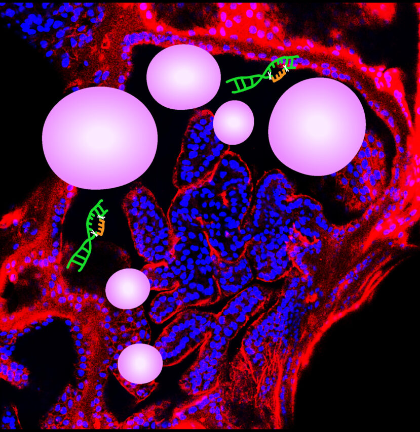
The Figure shows lenti-paired CRISPR-Cas9 gene editing for genetic disease ADPKD modeling in CD24+ derived adult human kidney tubuloids. Red color in Confocal image indicates CD24 staining, blue is DAPI, two cuts in DNA represent lenti-paired CRISPR-Cas9 gene editing in human PKD1 or PKD2 gene, white balloon-like structures represent ADPKD cysts.
From:
Yaoxian Xu and Rafael Kramann, Germany
Institute of Experimental Medicine and Systems Biology, Medical Clinic 2, Faculty of Medicine, RWTH Aachen University.
Author: Mayleen Laico, Philippines
Digital art
Info:
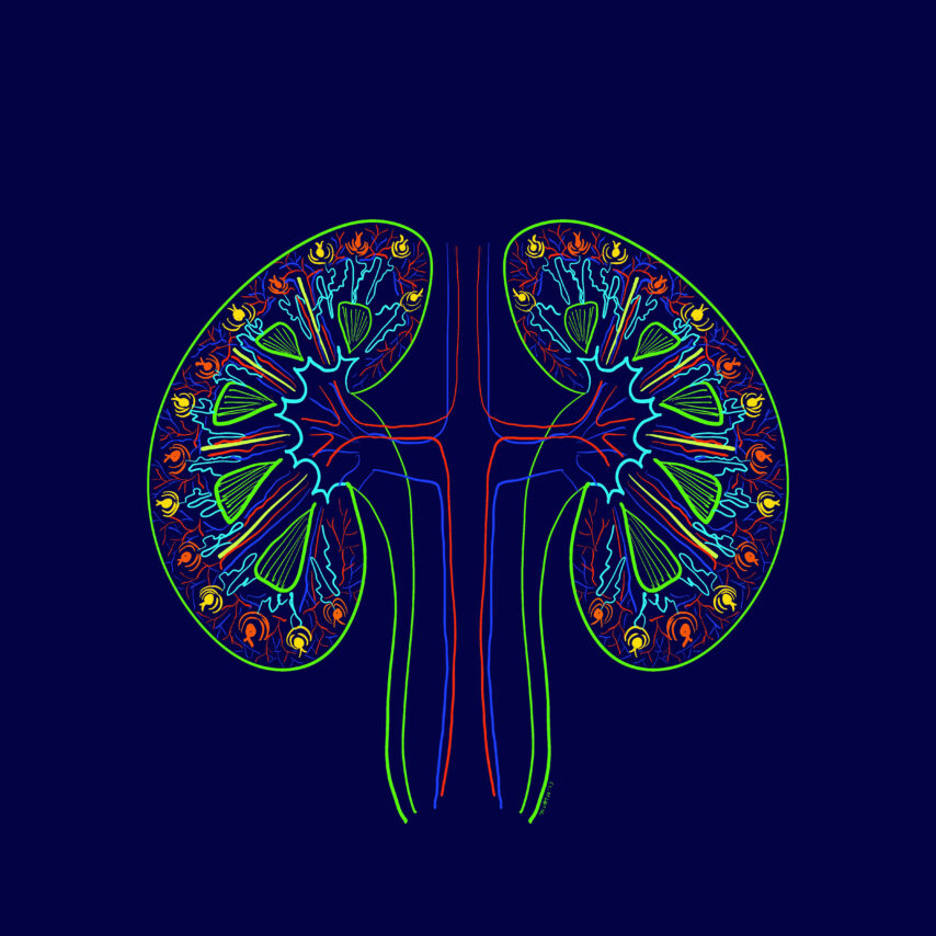
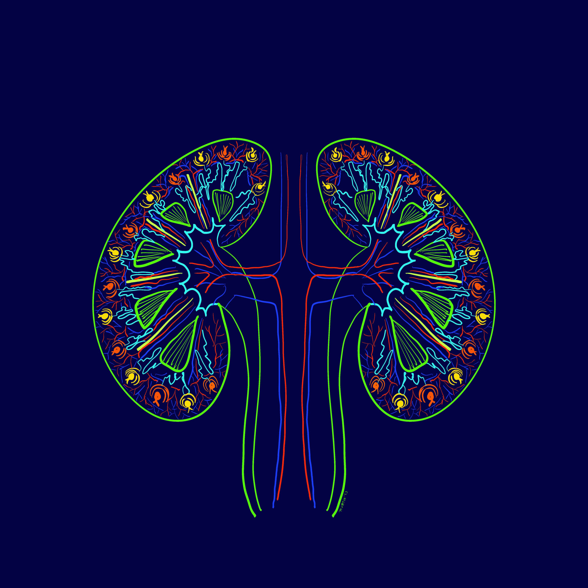
Organized Complexity shows the kidney’s complex function yet it is a very organized system
From:
Mayleen Laico, Philippines

Author: Xinyu Dong, USA
Drawing
Info:
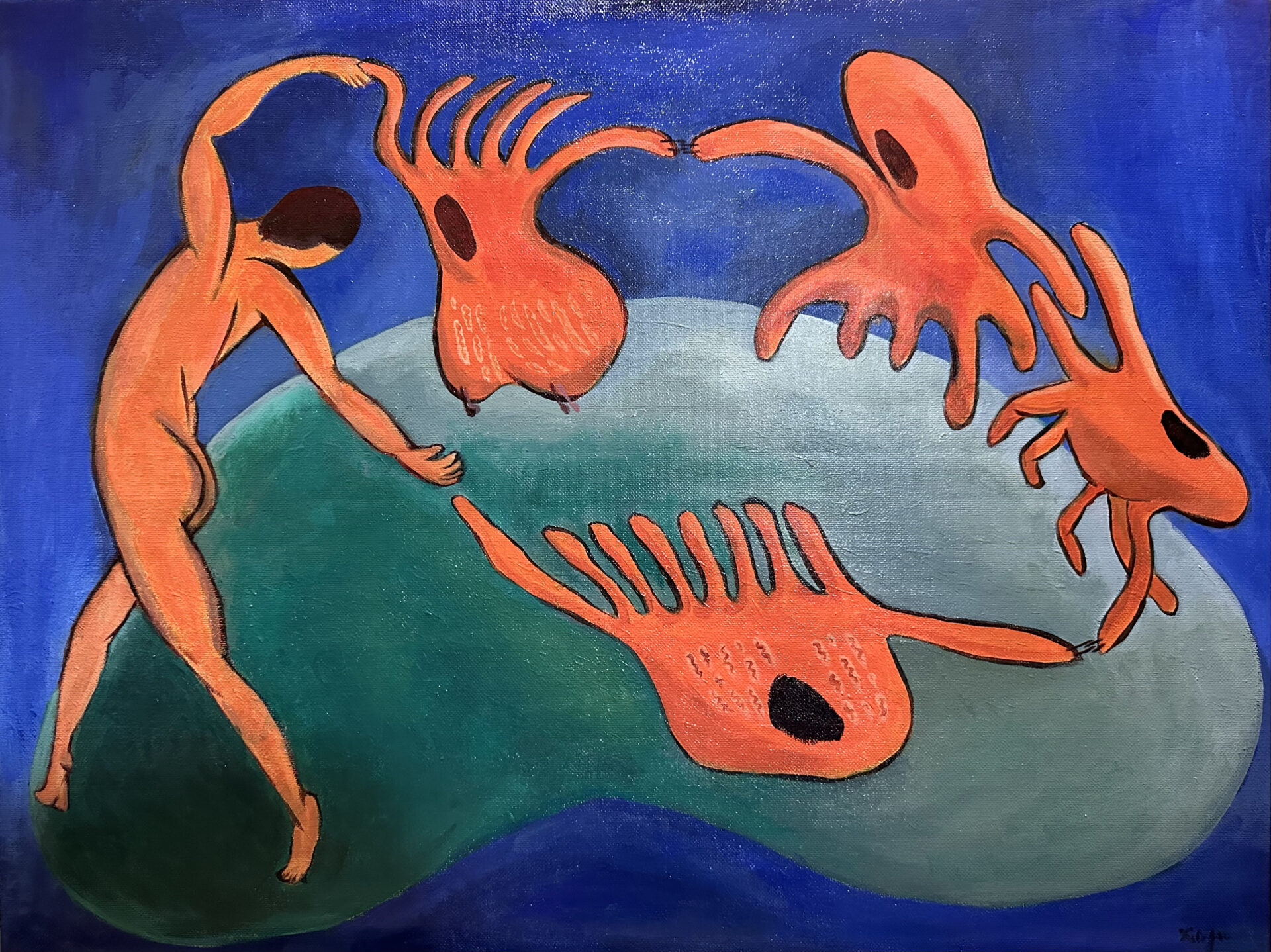
A nephrologist, immersed in passion, dances hand-in-hand with proximal tubule cells and podocytes. The dance unfolds on a green kidney, representing the earth, set against a blue cosmos background, symbolizing the nephrologist’s devotion to kidney research.
From:
Xinyu Dong, USA
Nephrology Department, Vanderbilt University Medical Center

Author: Sara Gjeci | Meike N. Leiske | Adrian T. Press, Germany
Advanced microscopy
Info:
Drug delivery system targeting kidney
From:
Sara Gjeci, Meike N. Leiske, Adrian T. Press
University of Jena and Bayreuth, Germany

Author: Marieta Theodorakopoulou, Greece
Histology
Info:
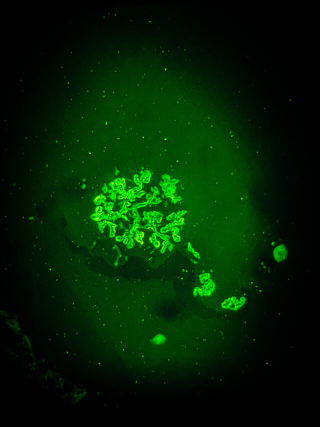
Glomerulus Galaxy – Immunofluorescence images stained for C3 in a patient with lupus nephritis
From:
Marieta Theodorakopoulou, Greece
1st Department of Nephrology, Hippokration Hospital, Aristotle University of Thessaloniki,

Author: Chenyu Li, Germany
Cells
Info:
Immunofluorescence images of cultured urine stem cells (stained for CD133 (red) and CD24 (green), collected from healthy human urine.
From:
Chenyu Li, Germany
Author: Ralph Mohty, USA
Urine & urine microscopy
Info:
Red blood cell cast indicating glomerulonephritis in a patient with AntiNeutrophil Cystoplasmic Antibody (ANCA) associated vasculitis
From:
Ralph Mohty, USA
MD MPH Stanford Nephrology

Author: Katharina Artinger, Austria
Advanced microscopy
NDT Cover February 2024
Info:
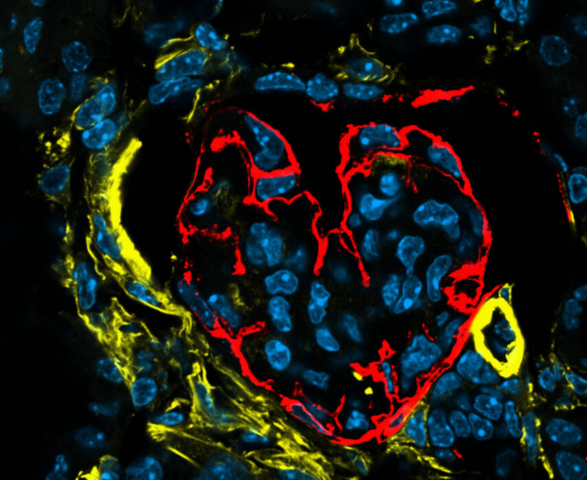
Love hurts – Heart-shaped glomerulus in a nephritic kidney
Podoplanin – red; Alpha smooth muscle actin – yellow; DAPI – blue.
From:
Katharina Artinger, Austria

Author: Anil Kumar Saxena, United Arab Emirates
Digital art
Info:
Focal Segmental Glomerulosclerosis
From:
Anil Kumar Saxena, United Arab Emirates
Chair, Nephrology Division Mediclinic Welcare Hospital Al Gahroud

Author: Barbara Mara Klinkhammer, Germany
Histology
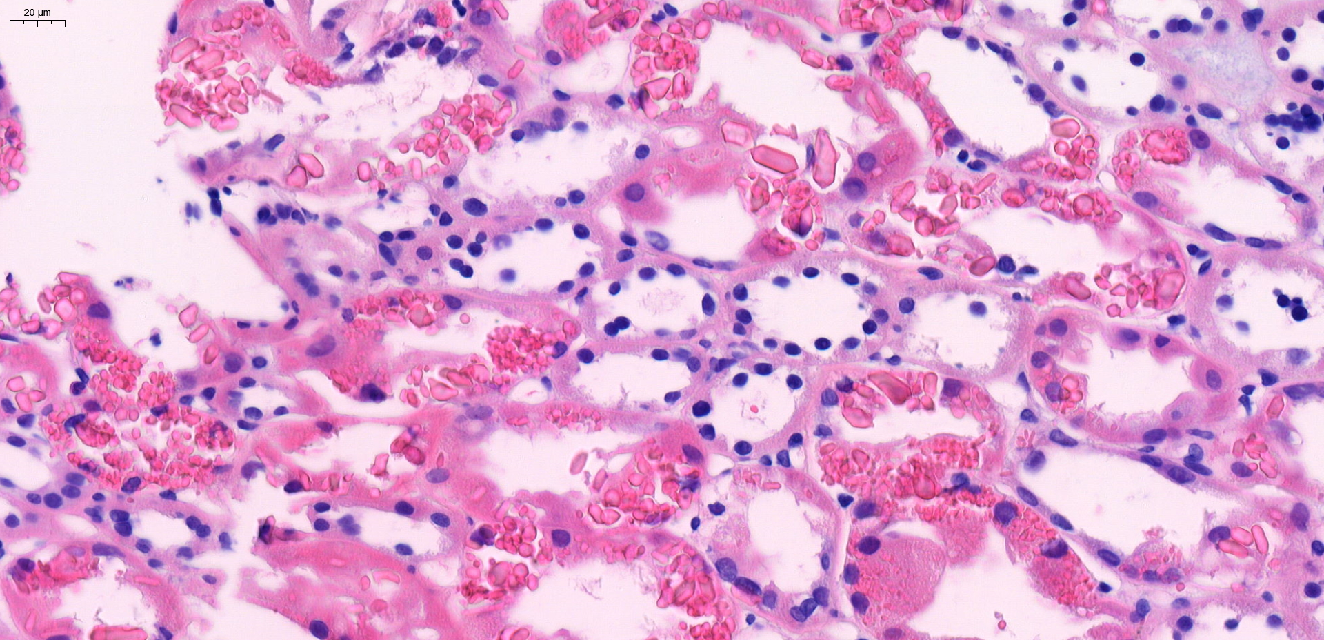
Info:
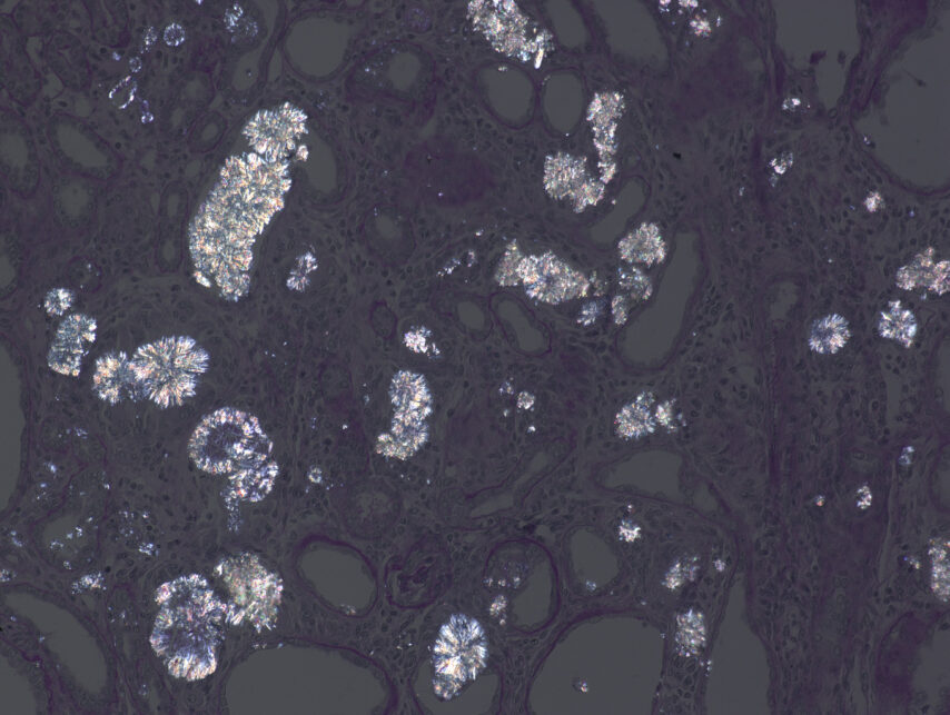
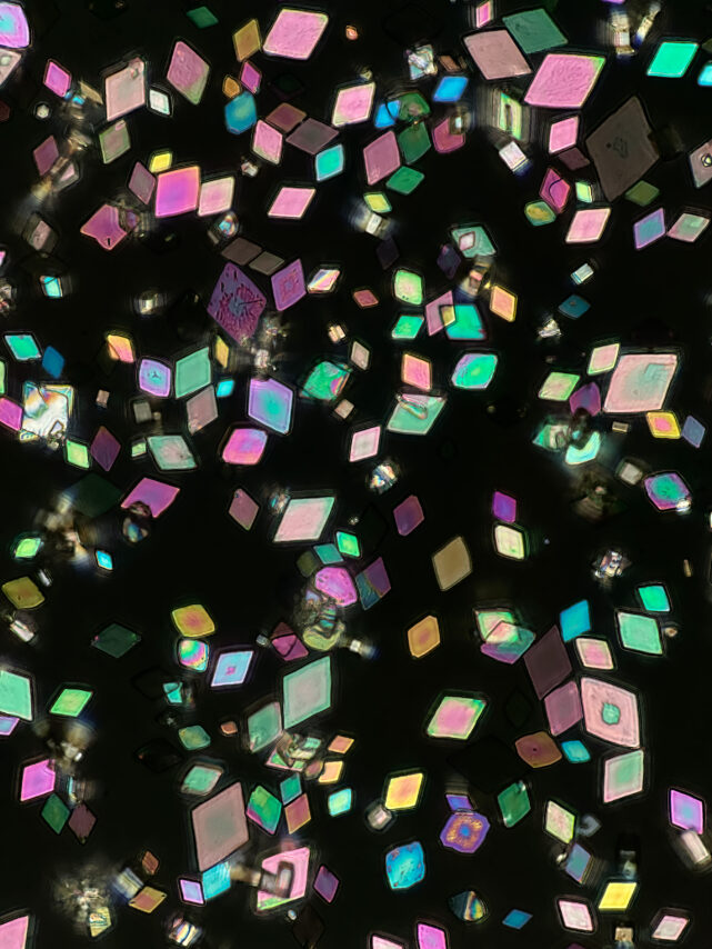
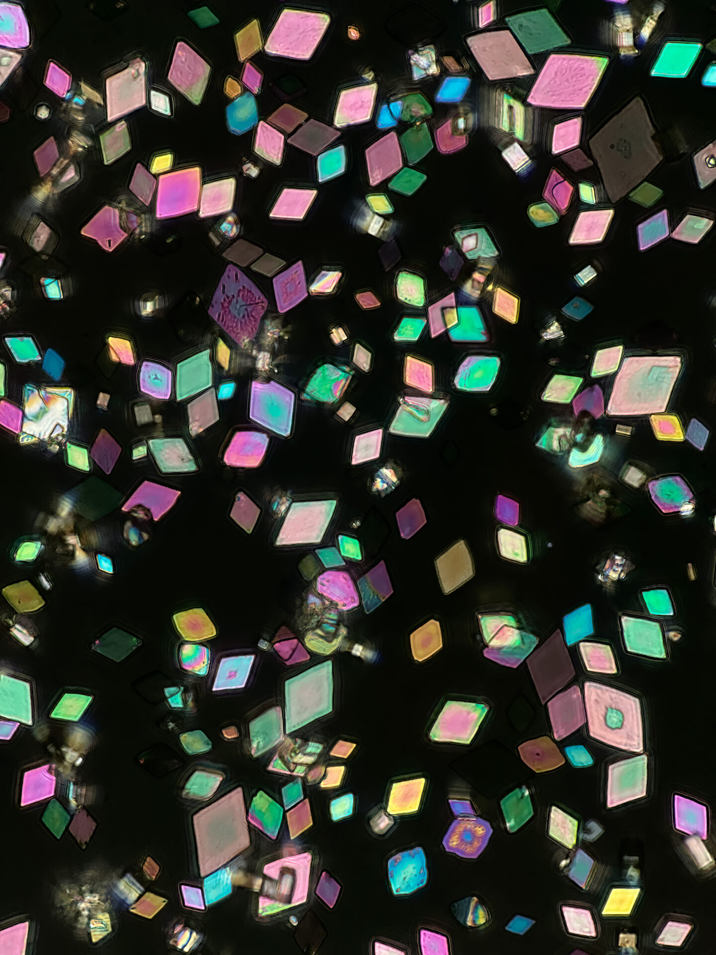
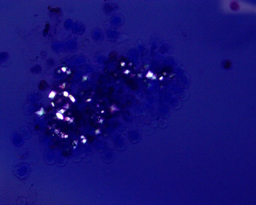
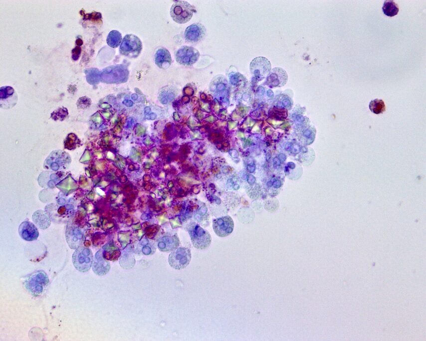
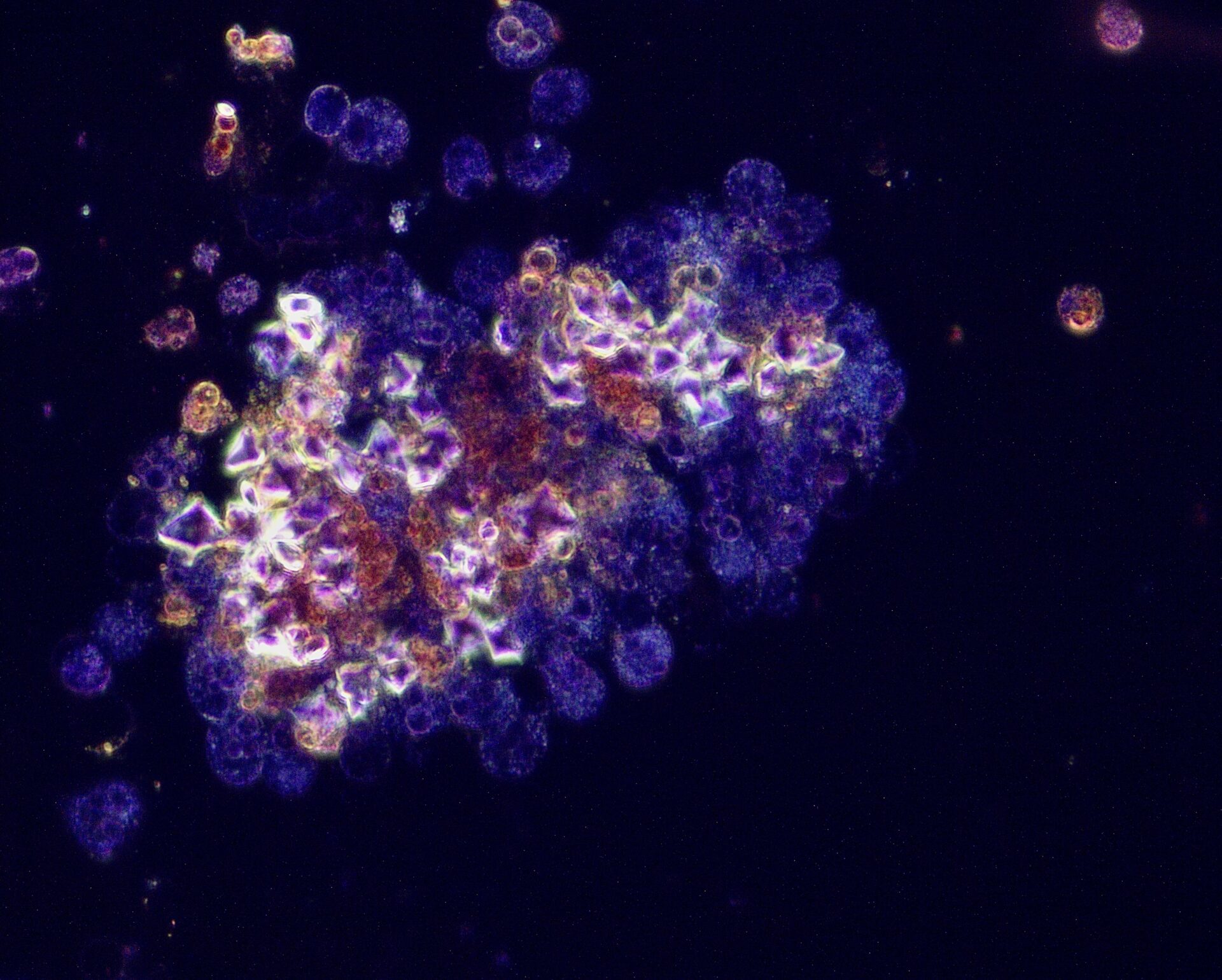
2,8-Dihydroxyadenine crystals in a rat kidney. PAS stain under polarized light.
From:
Barbara Mara Klinkhammer, Germany

Author: Sara Gjeci | Meike N. Leiske | Adrian T. Press, Germany
Imaging
Info:
Drug delivery system targeting kidney
From:
Sara Gjeci, Meike N. Leiske, Adrian T. Press
University of Jena and Bayreuth, Germany

Author: Monarch Shah, USA
Urine & urine microscopy
Info:
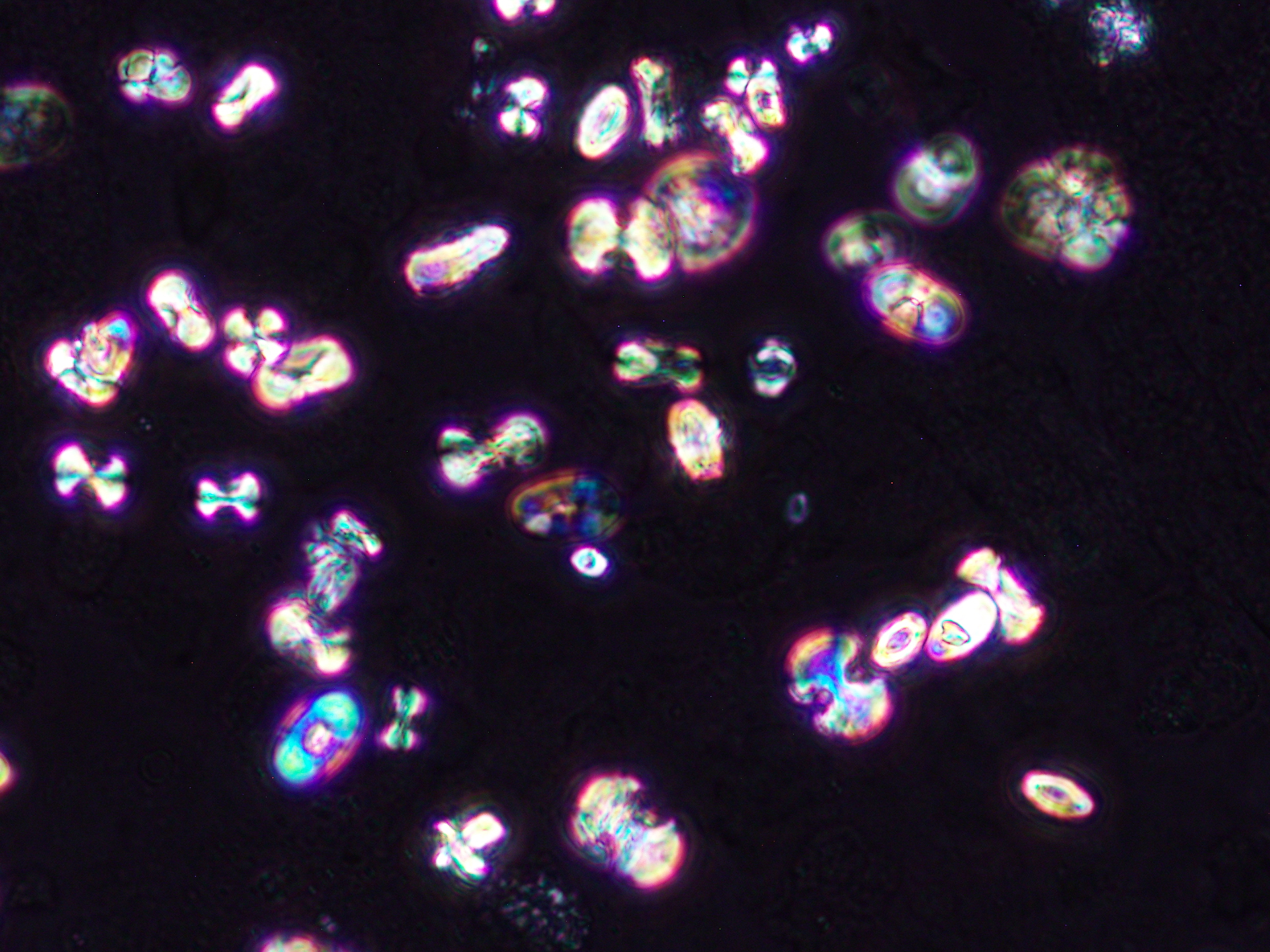
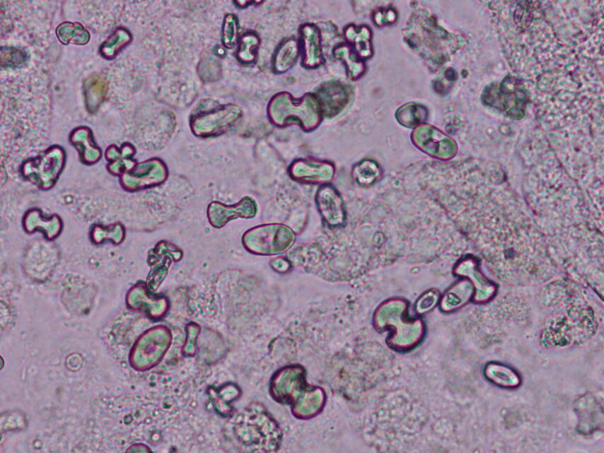
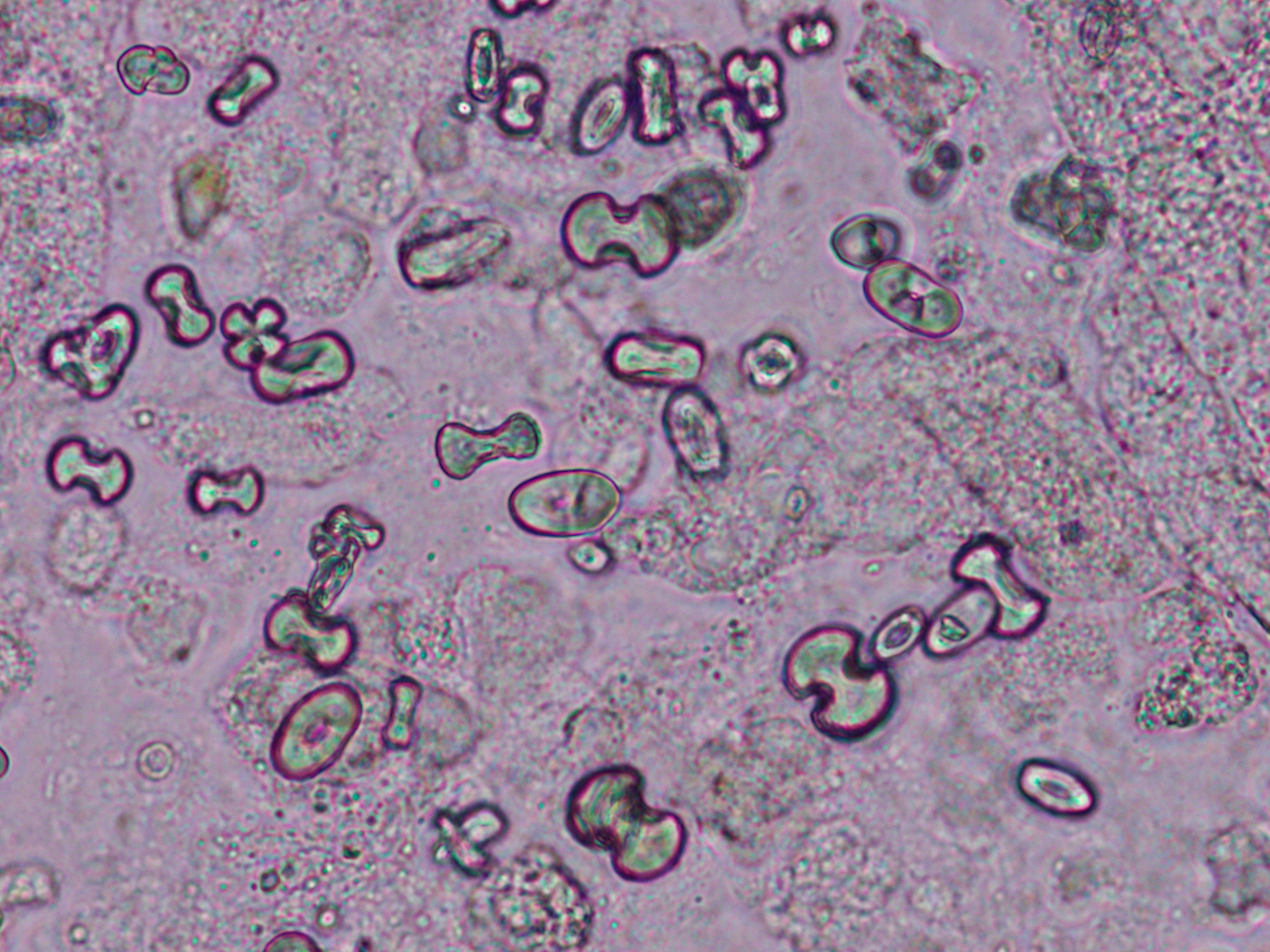
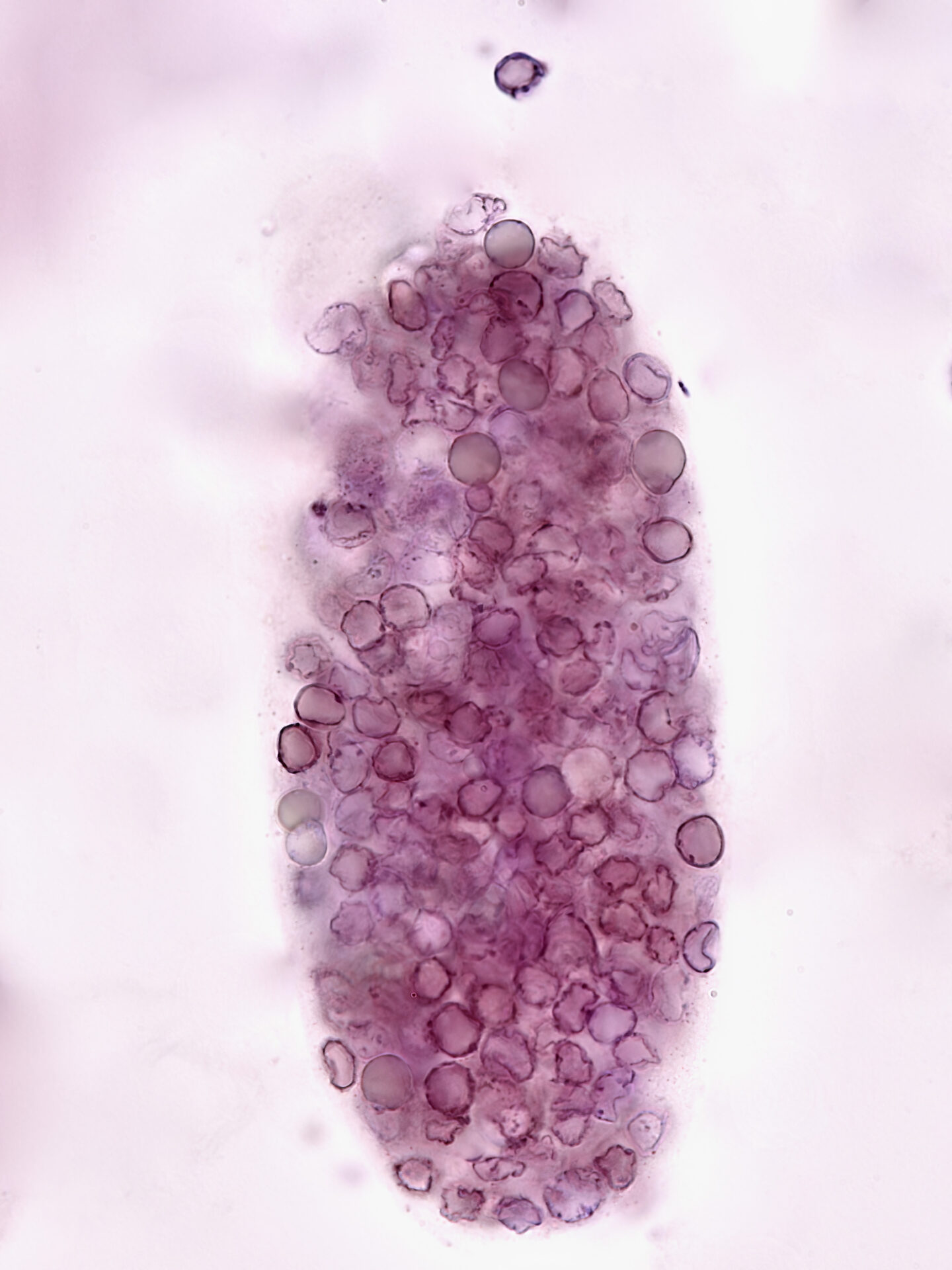
Calcium oxalate monohydrate crystals in a patient with acute kidney injury, many of the dumbbell-shaped type seen under phase contrast and polarized light (Original magnification x400).
From:
Monarch Shah,
Fellow, University of Virginia, USA
Author: Monarch Shah, USA
Urine & urine microscopy
Info:
Calcium oxalate monohydrate crystals in a patient with acute kidney injury, many of the dumbbell-shaped type seen under phase contrast and polarized light (Original magnification x400).
From:
Monarch Shah,
Fellow, University of Virginia, USA

Author: Ruben M Sandoval, USA
Advanced microscopy
Info:
Time projection showing filtration on the surface of a rat kidney acquired using intravital 2-photon microscopy.
A bolus of a small blue dextran appears first in the lumen of the Bowman’s space, proximal tubule lumen, then finally in the cortical collecting duct lumen.
Projection is 300 frames, close to 4 minutes.
From:
Ruben M Sandoval, USA
Indiana Center for Biological Microscopy
Author: Ruben M Sandoval, USA
Advanced microscopy
NDT Cover December 2023
Info:
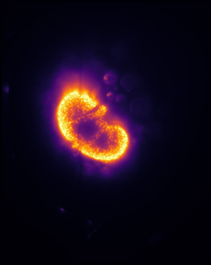
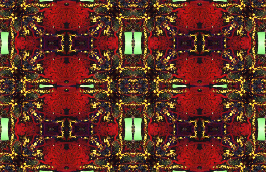
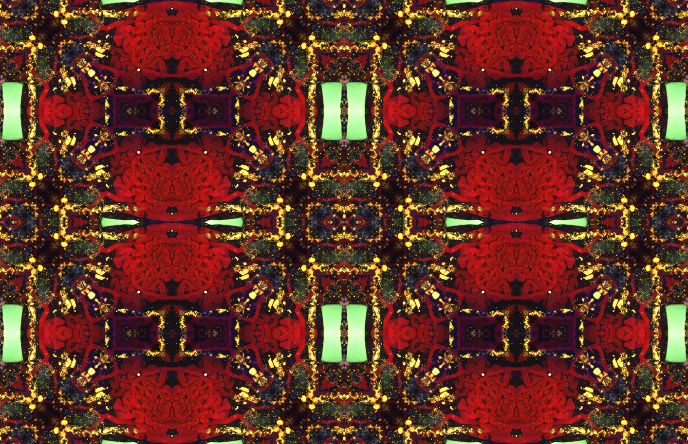
A chronic kidney disease rat model showing the distribution of albumin (red) acquired in 3D using intravital 2-photon microscopy.
Fluorescent cast material (green) can be seen obstructing the lumen of a distal tubule. The original image was processed to generate a tessellation with seamless borders and natural symmetry.
From:
Ruben M Sandoval, USA
Indiana Center for Biological Microscopy

Author: Hélène Fank, Belgium
Electron microscopy
Info:
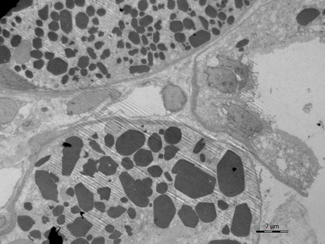
Electron microscopy confirmed the electron-dense crystalline structures within cytoplasm of proximal tubular epithelial cells
From:
Hélène Fank, Belgium
MD

Author: Hélène Fank, Belgium
Histology
Info:
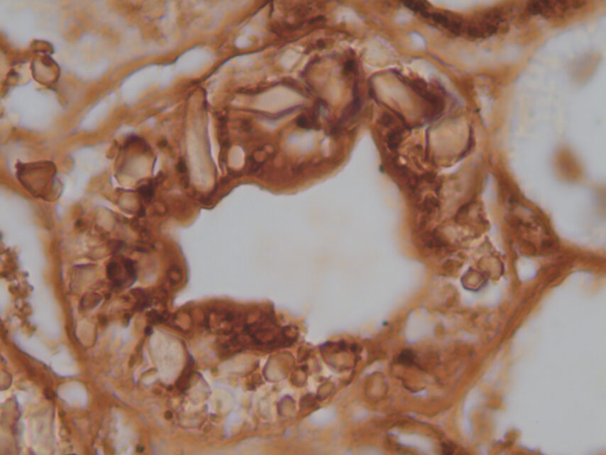
Immunohistochemistry for kappa light chains was strongly positive within these crystals (b, white arrow)
From:
Hélène Fank, Belgium
MD

Author: Hélène Fank, Belgium
Histology
Info:
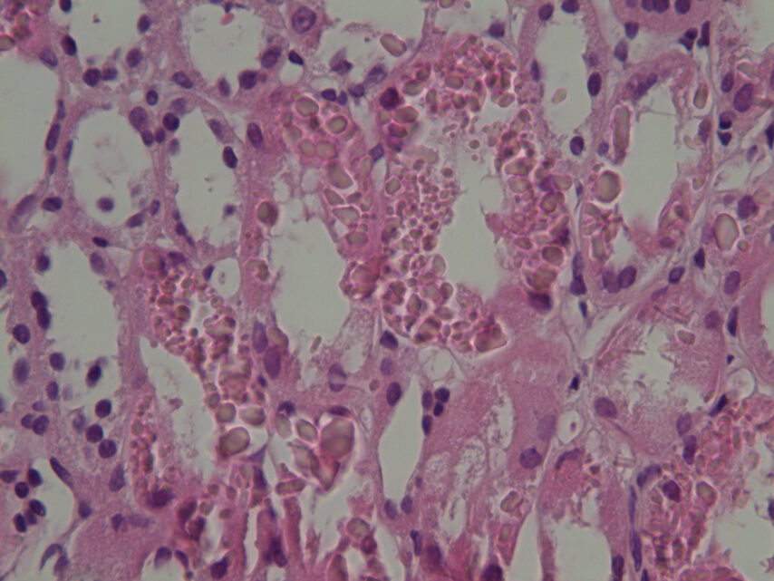
Light microscopy (Hematoxylin x400) showing intracellular crystalline inclusions within proximal tubular epithelial cells (a, black arrow)
From:
Hélène Fank, Belgium
MD
Author: Mayleen Laico, Philippines
Digital art
NDT Cover November 2023
Info:
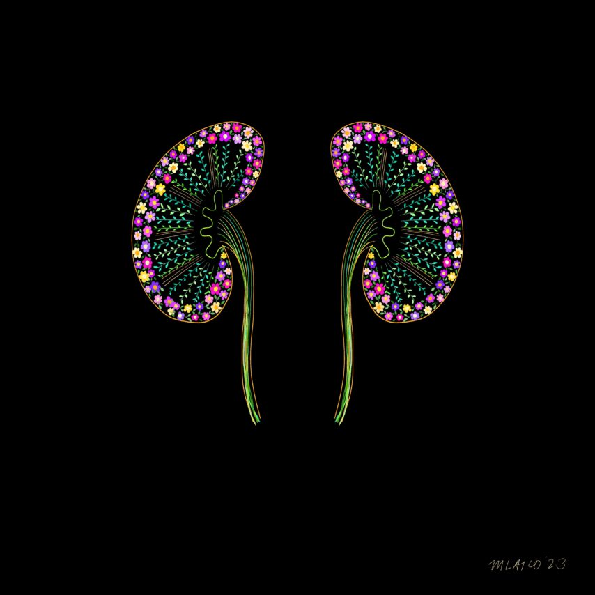
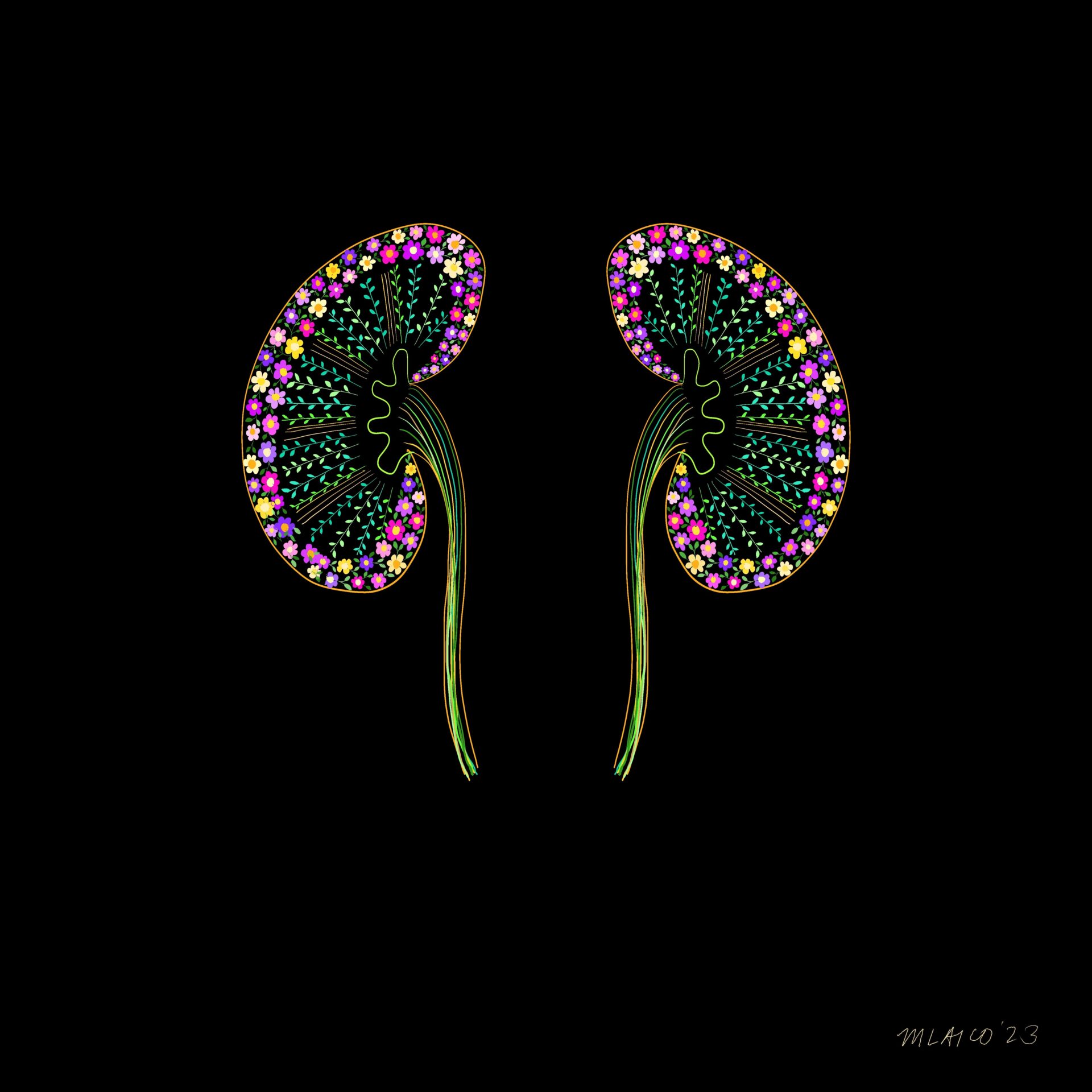
The Greening of Nephrology
From:
Mayleen Laico, Philippines
Author: J. Romero Tafoya, México
Urine & urine microscopy
Info:
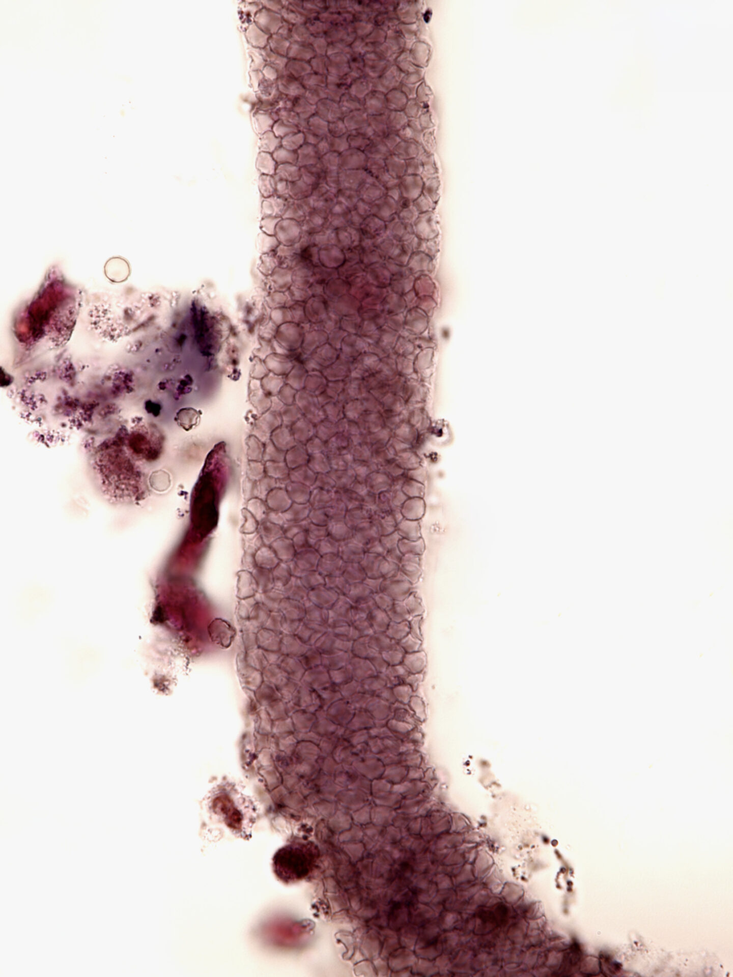
Red blood cell cast – brightfield with Sternheimer – Malbin stain
From:
J. Romero Tafoya, México
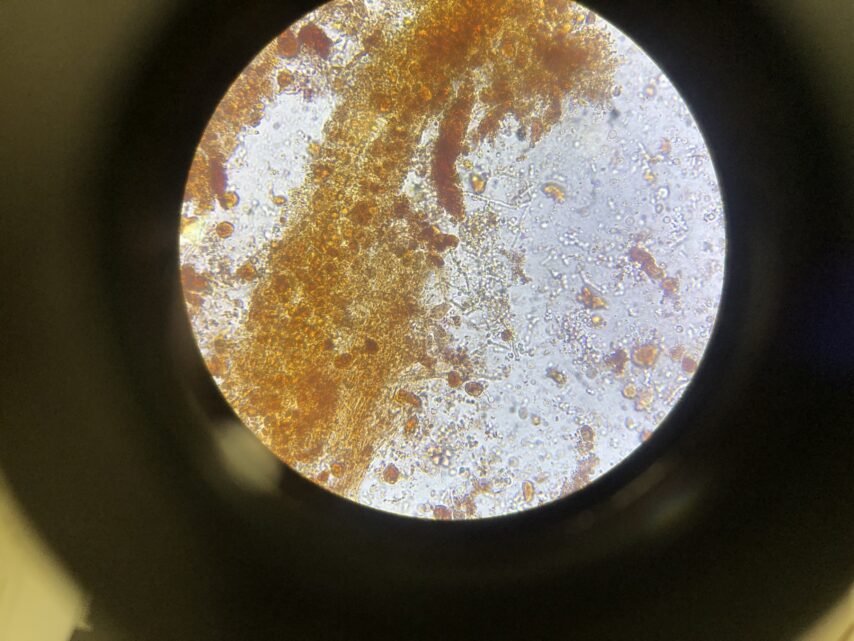

Author: J. Romero Tafoya, México
Urine & urine microscopy
Info:
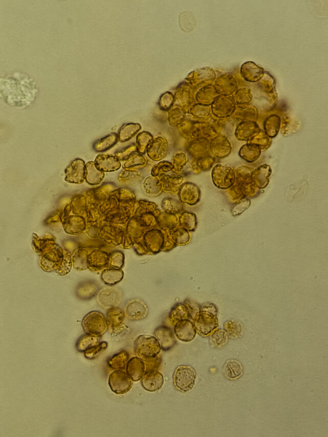
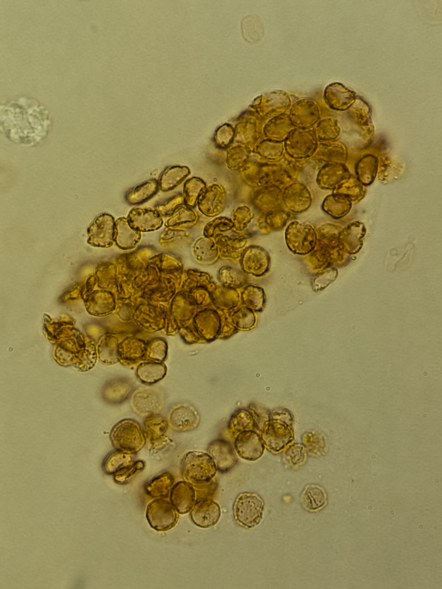
Bile cast and hyphae – brightfield with Sternheimer – Malbin stain
From:
J. Romero Tafoya, México

Author: J. Romero Tafoya, México
Urine & urine microscopy
Info:
Red blood cell cast- brightfield with Sternheimer- Malbin stain
From:
J. Romero Tafoya, México
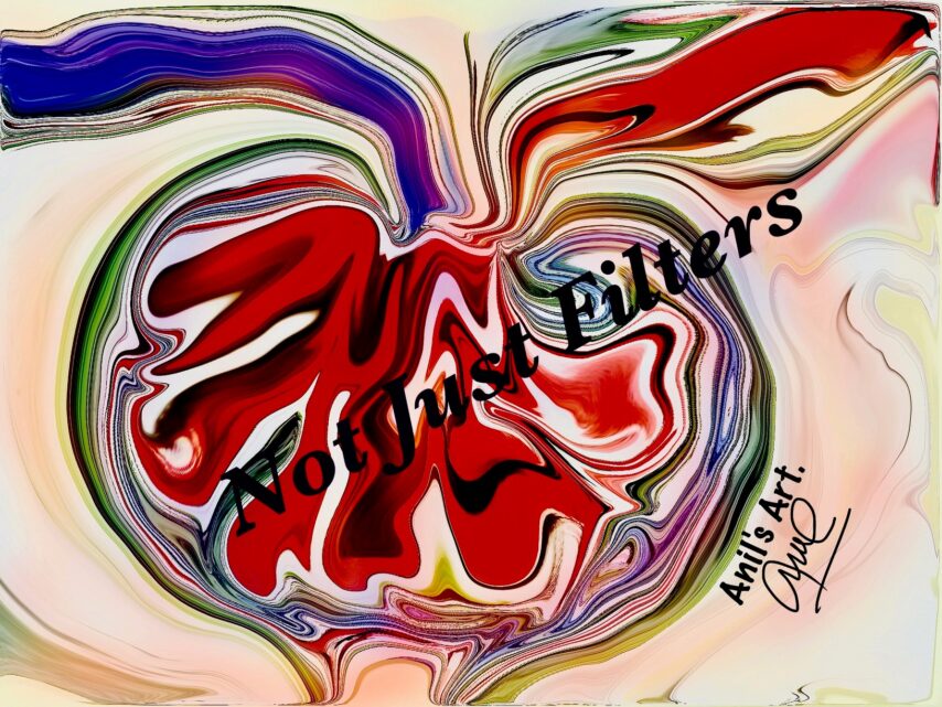
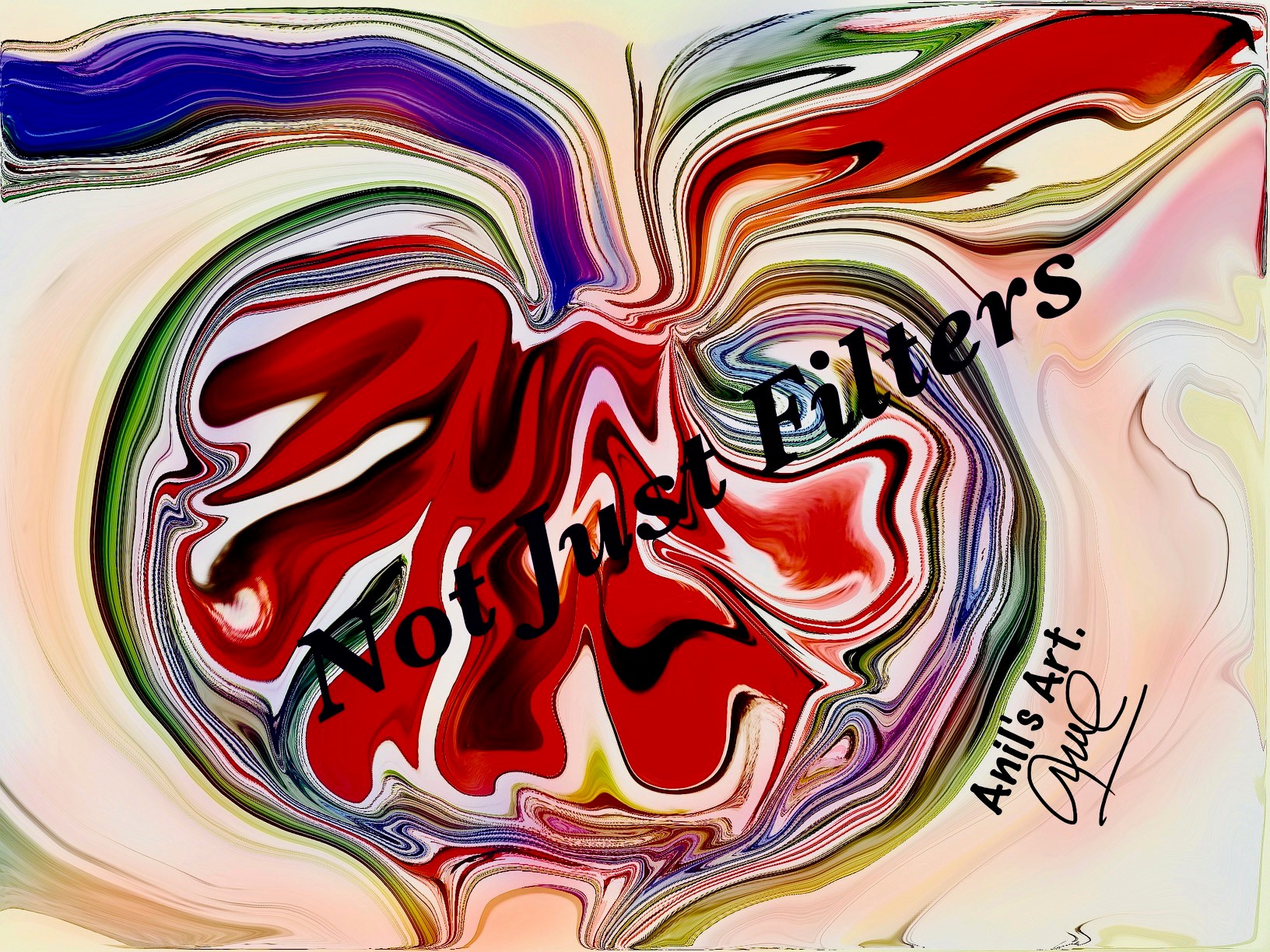
Author: Anil Saxena
Digital art
Info:
The Glomerulus ( Not Just Filters)
From:
Anil Saxena, United Arab Emirates
MD, FRCP, FASN Chair,
Nephrology Division Medical Director

Author: Reynaldo Noriega Flores, México
Drawing
Info:
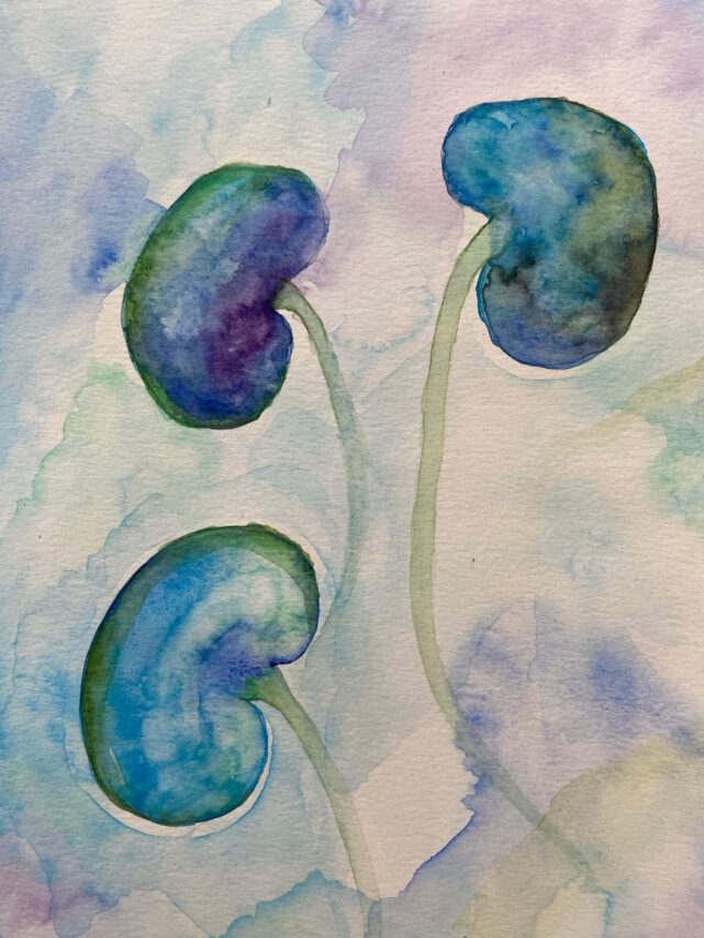
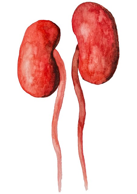
Kidney Transplant
Watercolour painting
From:
Reynaldo Noriega Flores, México

Author: Pierre-Emmanuel N'Guetta and Lori O'Brien, USA
Advanced microscopy
NDT Cover October 2023
Info:
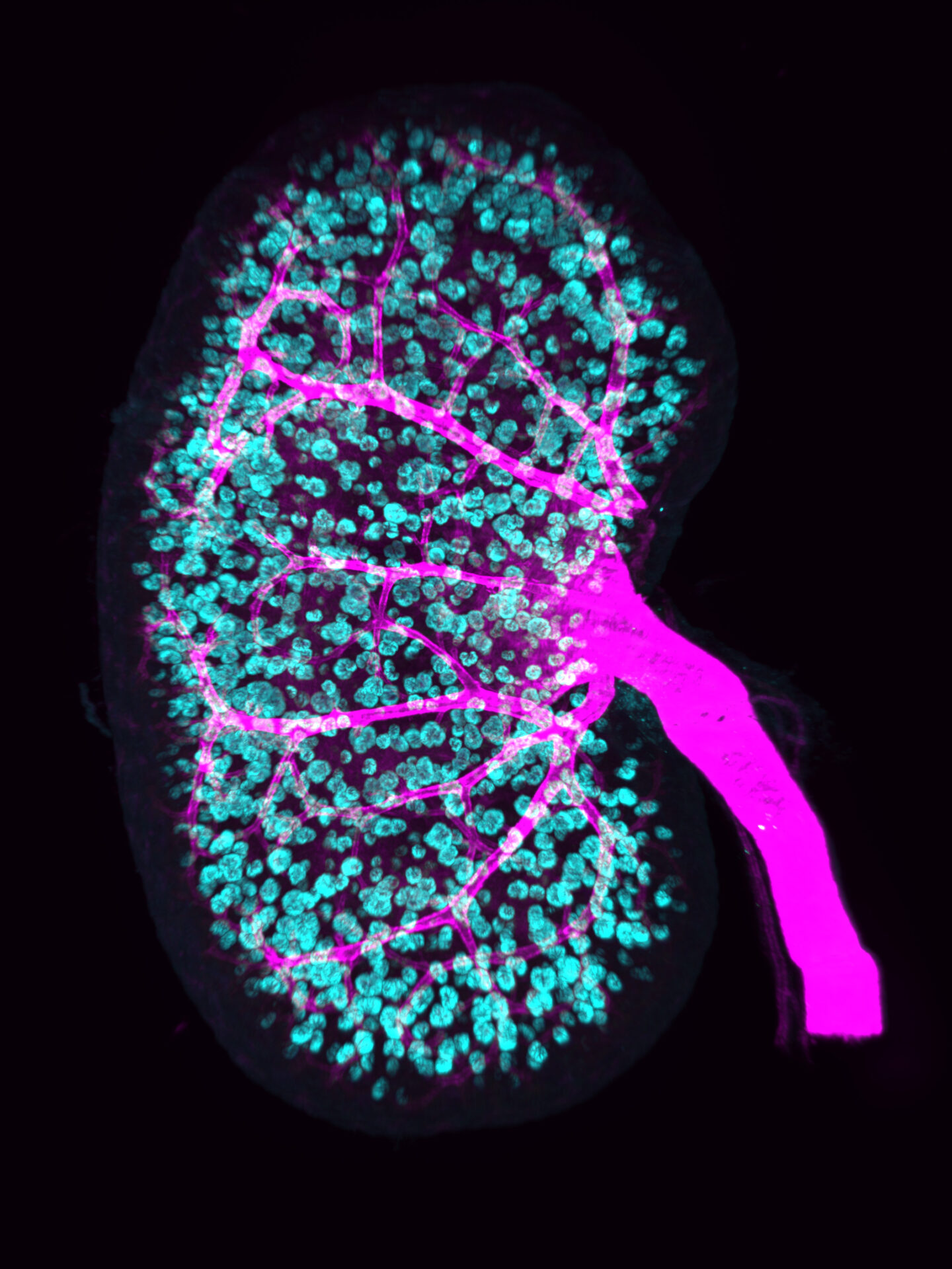
This image depicts a postnatal mouse kidney immunostained for the renal arterial tree with alpha-Smooth Muscle Actin (SMA, magenta) and glomeruli with podocin (Nphs2, cyan). Kidney was imaged with a LaVision Ultramicroscope II LightSheet using an Olympus MVPLAPO 2x/0.5 objective, a 2x Zoom, Sheet NA of 0.23, a Z step of 2.0µm and image pixel size of 1.54µm/pixel.
From:
Pierre-Emmanuel N’Guetta, USA
Lori O’Brien, USA
PhD, Assistant Professor

Author: Jay R. Seltzer, USA
Urine & urine microscopy
Info:
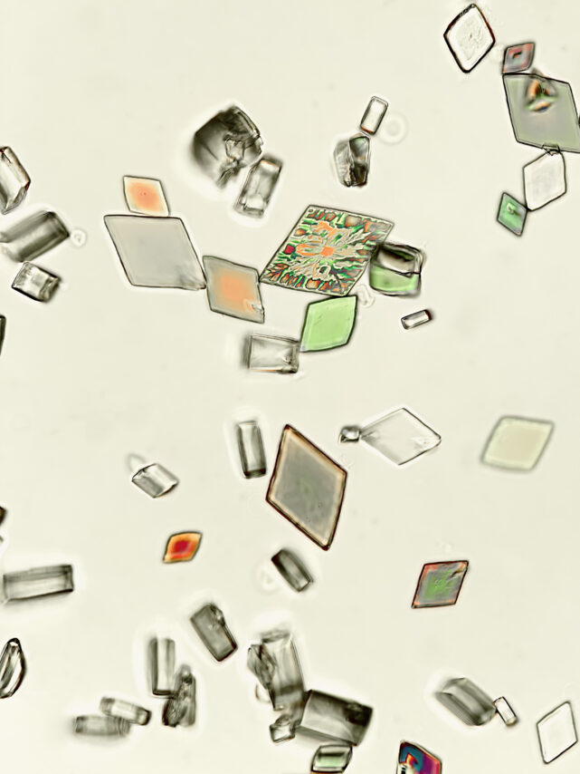
Uric acid crystals, under partial polarization – original magnification x400 – from patient with marked volume depletion and AKI
From:
Jay R. Seltzer, USA
MD, Chief of Nephrology

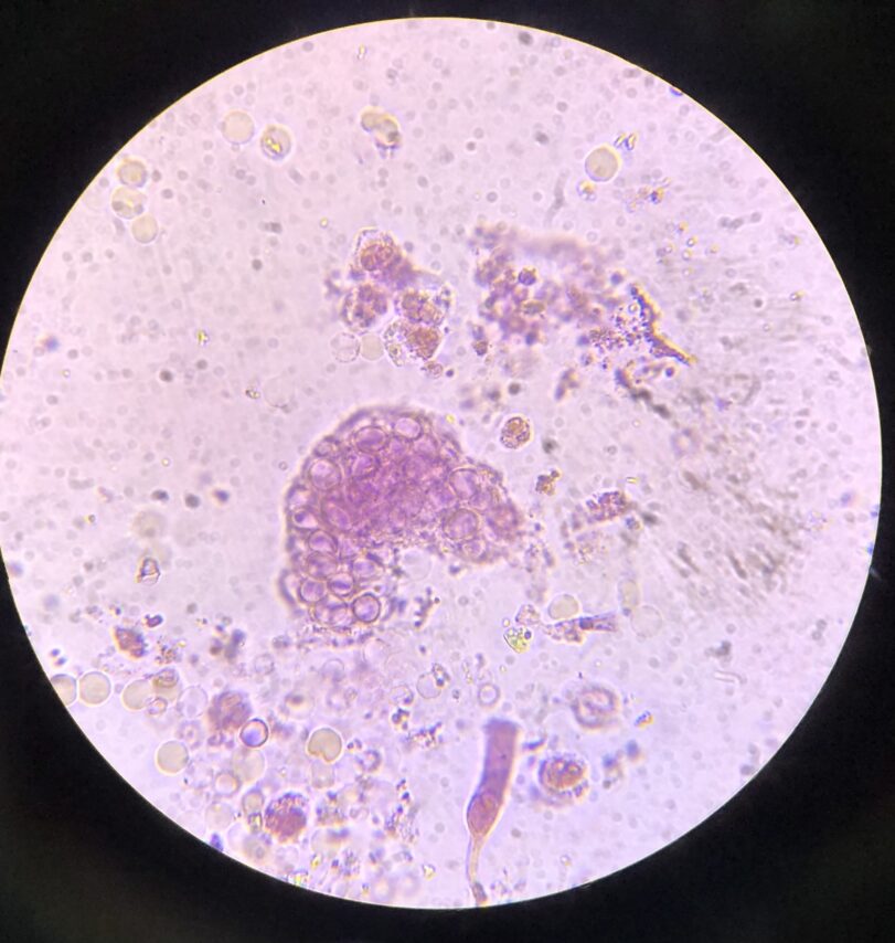
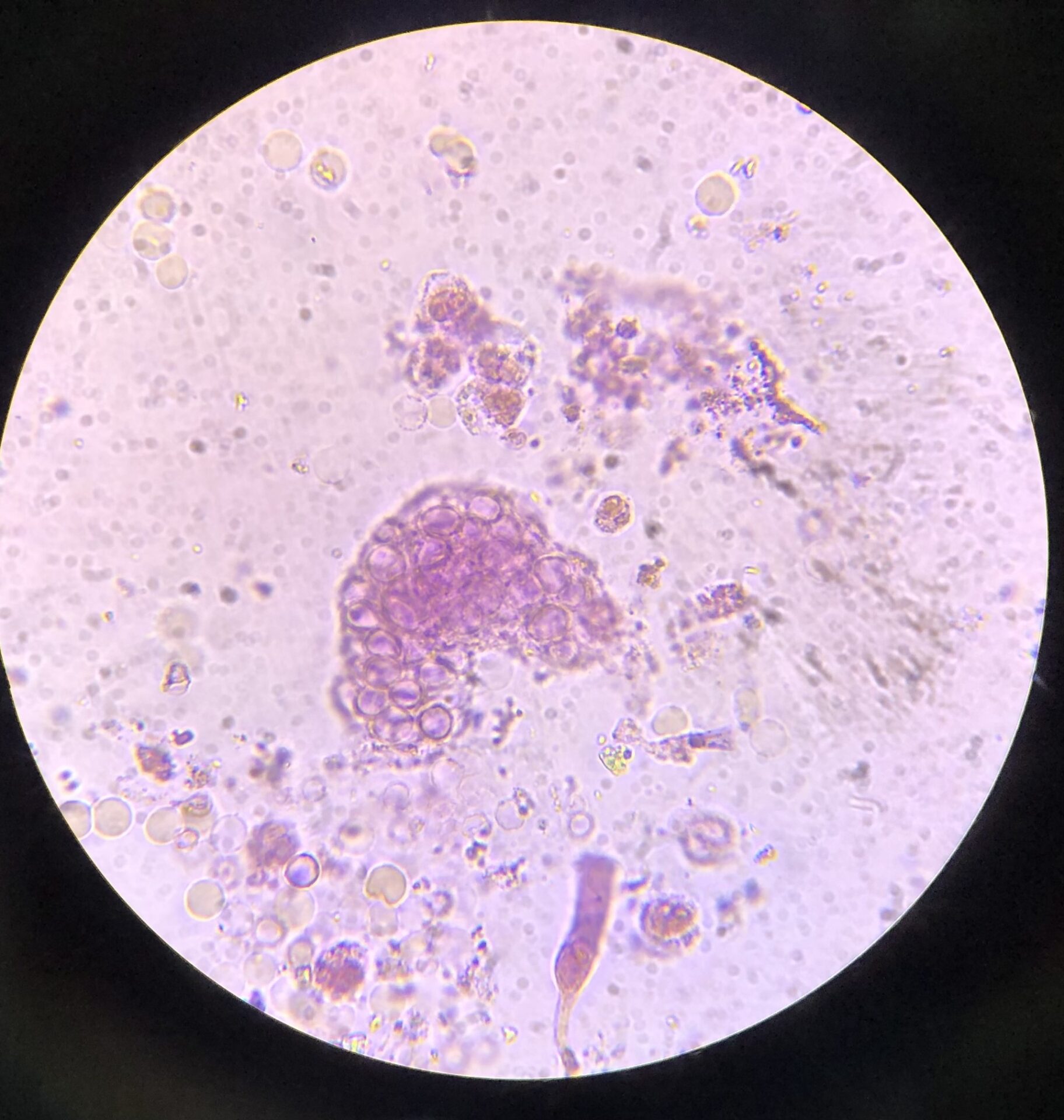
Author: Jay R. Seltzer, USA
Urine & urine microscopy
Info:
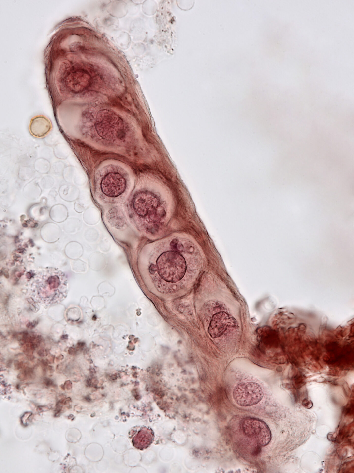
Renal tubular epithelial cell cast – brightfield with prolonged Sternheimer-Malbin staining – original magnification x1000 – from patient with bacterial endocarditis, acute kidney injury, and infection related glomerulonephritis
From:
Jay R. Seltzer, USA
MD, Chief of Nephrology
Author: Jay R. Seltzer, USA
Urine & urine microscopy
NDT Cover September 2023
Info:
Uric acid crystals in urine, under fully polarized light – original magnification x400 – from patient with marked volume depletion and AKI
From:
Jay R. Seltzer, USA
MD, Chief of Nephrology

Author: Jay R. Seltzer, USA
Urine & urine microscopy
Info:
Red blood cell cast – brightfield with Sternheimer-Malbin stain – original magnification x1000 – from patient with hydralazine-induced vasculitis
From:
Jay R. Seltzer, USA
MD, Chief of Nephrology
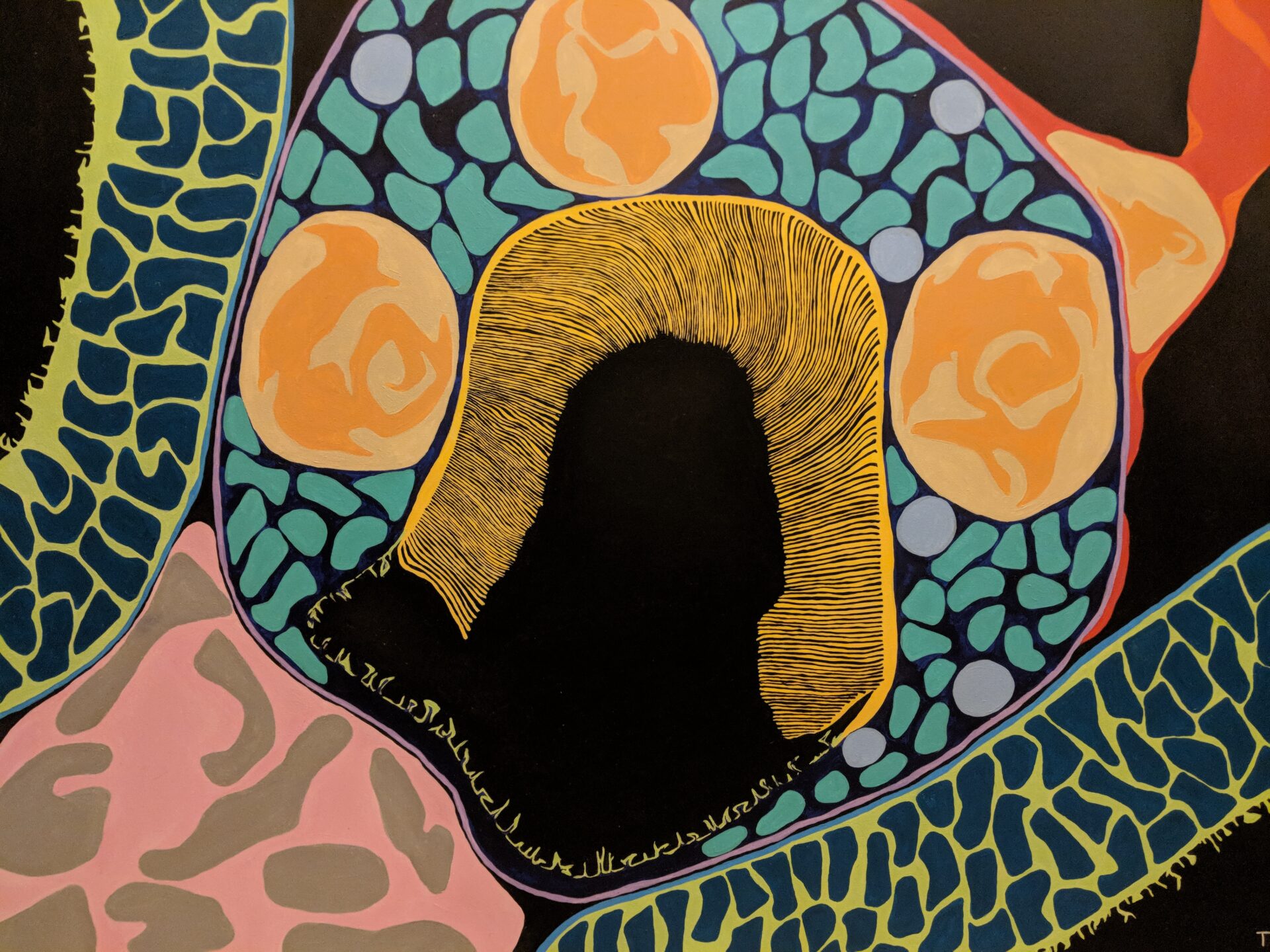
Author: Tiffany Caza, USA
Drawing
NDT Cover July 2023
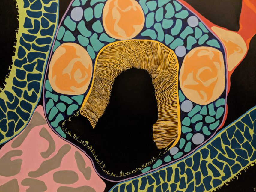
Info:
This image demonstrates an abstract view of the transition between a proximal tubule and the thick descending limb of the loop of Henle
From:
Tiffany Caza, USA
MD/PhD, a nephropathologist at Arkana Laboratories

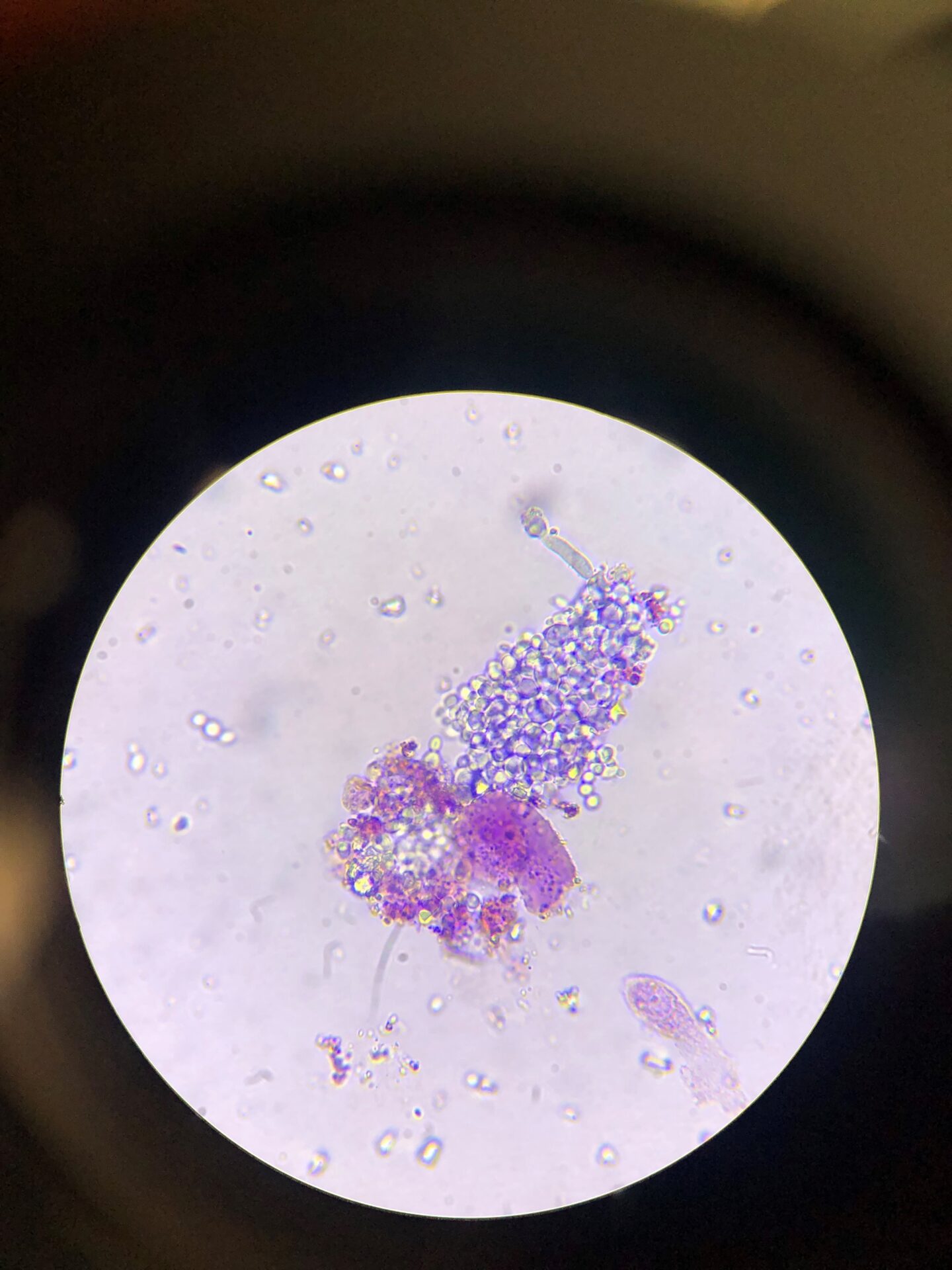
Author: Swetha Kanduri, USA
Urine & urine microscopy
Info:
B: Bright field, dark field and phase contrast image of cluster of calcium oxalate dihydrate crystals surrounding leukocytes
From:
Swetha Kanduri, USA
MD, Assistant Professor of Medicine
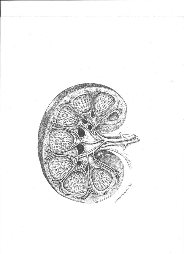

Author: Maia Celeste Arbatin, Philippines
Drawing
Info:
Pen and ink sketch
From:
Maia Celeste Arbatin, Philippines
MD FPCP FPSN (#nephgirlsketches)
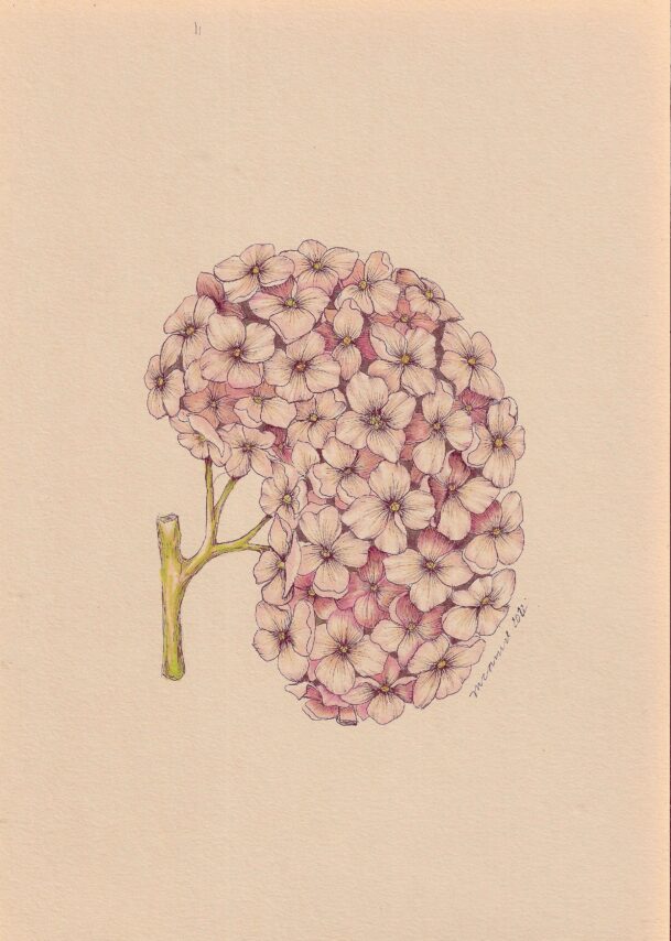
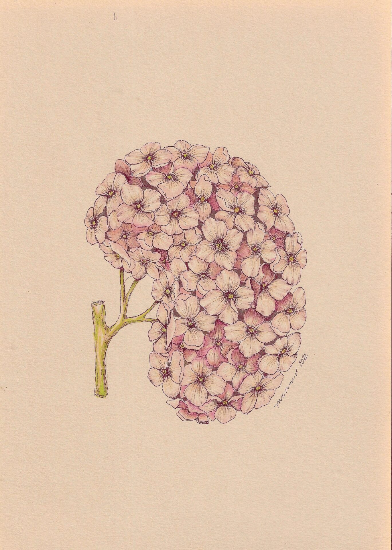
Author: Maia Celeste Arbatin, Philippines
Drawing
Info:
Hydrangea Kidney
From:
Maia Celeste Arbatin, Philippines
MD FPCP FPSN (#nephgirlsketches)


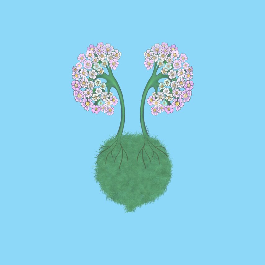
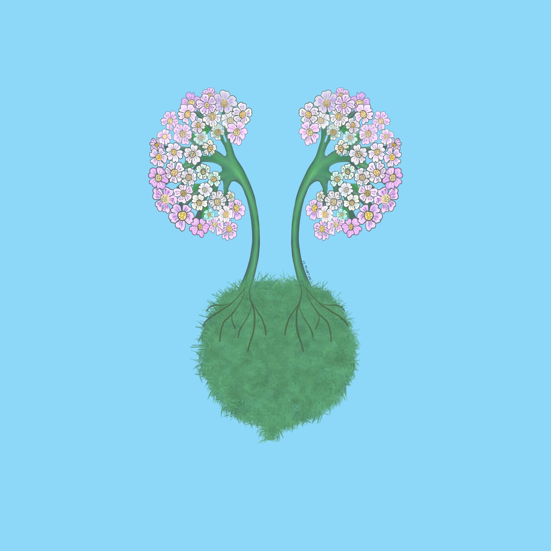
Author: Jeanine Verbeke, Belgium
Drawing
Info:
Kidney Trees
From:
Jeanine Verbeke, Belgium

Author: Dominique Couck, Belgium
Drawing
NDT Cover August 2023
Info:
Drawing for a family gift
From:
Dominique Couck, Belgium

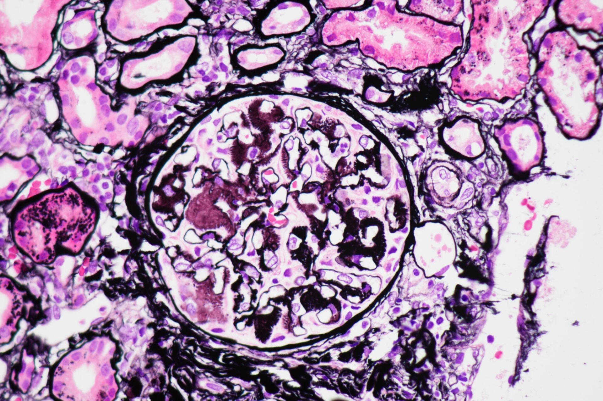
Author: Boonyarit Cheunsuchon, Thailand
Histology
Info:
Eyelash sign in glomerular amyloidosis
From:
Boonyarit Cheunsuchon, Thailand
MD, Associate professor
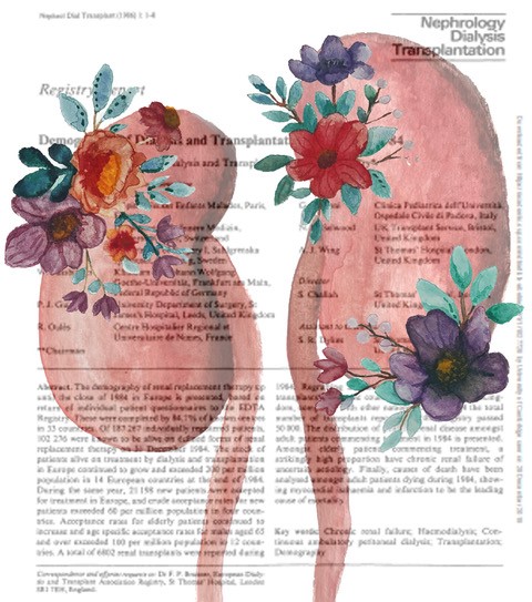
Author: Sonja Djudjaj, Germany
Digital art
Info:
NDT history
From:
Sonja Djudjaj, Germany
PhD, Assistant Professor
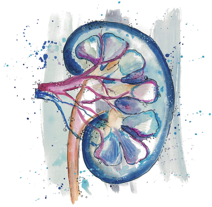
Author: Sonja Djudjaj, Germany
Drawing
Info:
Water color kidney
From:
Sonja Djudjaj, Germany
PhD, Assistant Professor
Author: Mayleen Laico, Philippines
Digital art
Info:
Giving hope
From:
Mayleen Laico, Philippines
MD

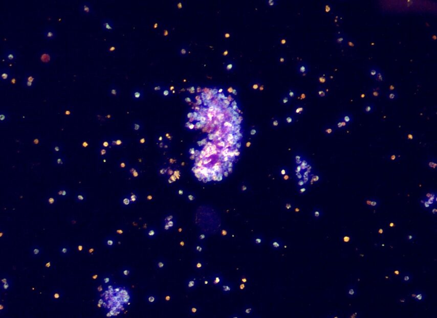
Author: Maria Lucia Angelotti, Italy
Advanced microscopy
Info:
Representative image obtained by STED super-resolution microscopy, showing strong positivity for IgA deposits (orange) and a partial mild effacement of foot processes (stained with nephrin, cyan) in a biopsy of IgA nephropathy patient. The biopsy specimen underwent optical tissue clearing before staining.
From:
Maria Lucia Angelotti, Italy
PhD

Author: Miriam Buhl, Germany
Electron microscopy
Info:
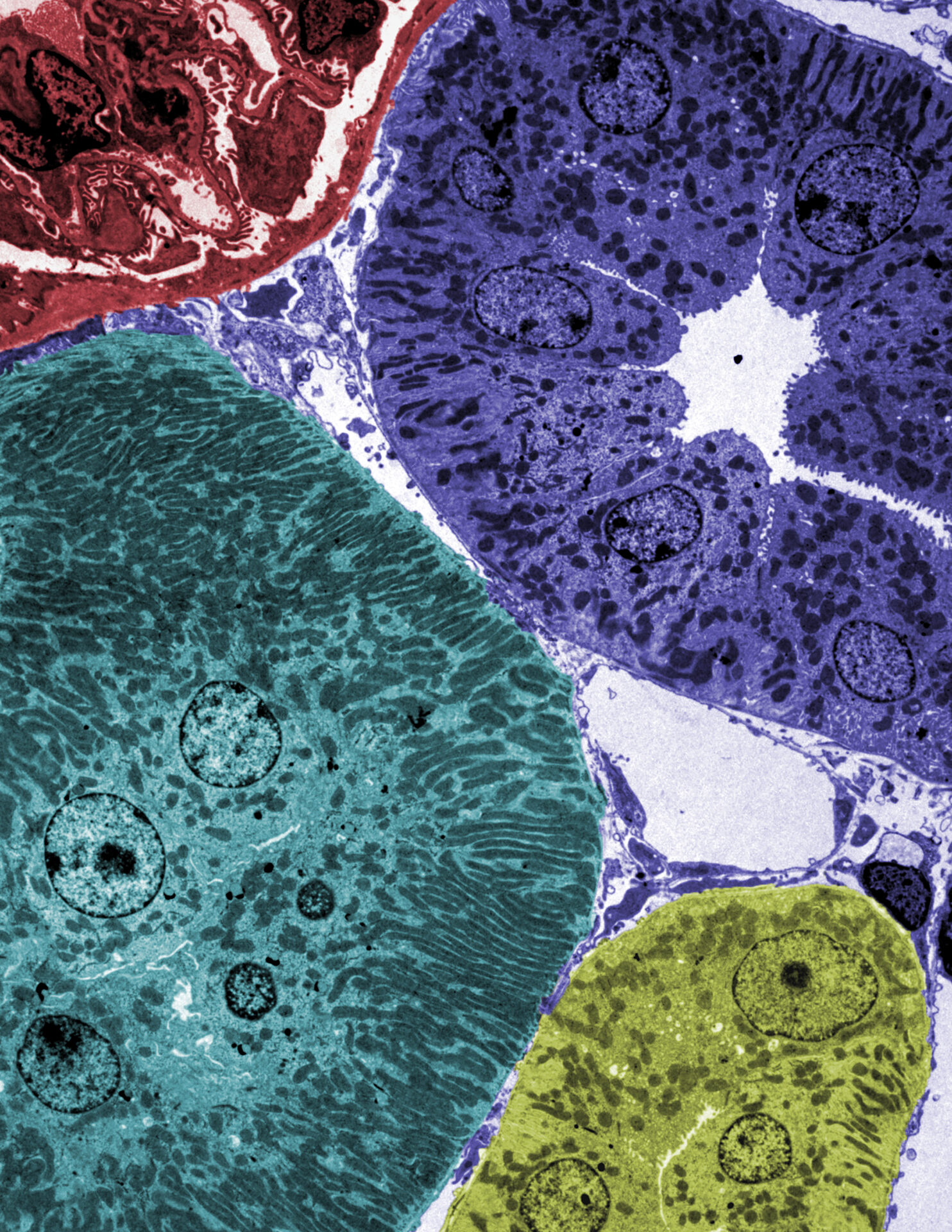
Colorized transmission electron microscopy image of murine renal kidney cortex. Individual tubular segments are colored in green, blue and yellow, while a glomerular section is colored in red.
From:
Miriam Buhl, Germany
PhD

Author: Swetha Kanduri, USA
Urine & urine microscopy
Info:
Phase contrast image of cluster of calcium oxalate dihydrate crystals surrounding leukocytes
From:
Swetha Kanduri, USA
MD, Assistant Professor of Medicine

Author: Swetha Kanduri, USA
Urine & urine microscopy
Info:
Bright field image of cluster of calcium oxalate dihydrate crystals surrounding leukocytes
From:
Swetha Kanduri, USA
MD, Assistant Professor of Medicine

Author: Swetha Kanduri, USA
Urine & urine microscopy
Info:
Dark field image of cluster of calcium oxalate dihydrate crystals surrounding leukocytes
From:
Swetha Kanduri, USA
MD, Assistant Professor of Medicine

Author: Jay R. Seltzer, USA
Urine & urine microscopy
Info:
Red blood cell cast – brightfield with Sternheimer-Malbin stain – original magnification x1000 – from patient with anti-glomerular basement membrane disease
From:
Jay R. Seltzer, USA
MD, Chief of Nephrology

Author: Hélène Fank, Belgium
Histology
Info:
Secondary Toni-Debré-FANCONI syndrome – intracellular crystalline inclusions within proximal tubular epithelial cells
From:
Hélène Fank, Belgium
MD
Co-authors:
Antoine Bouquegneau, Christophe Bovy and Orphal Colleye

Author: Boonyarit Cheunsuchon, Thailand
Histology
Info:
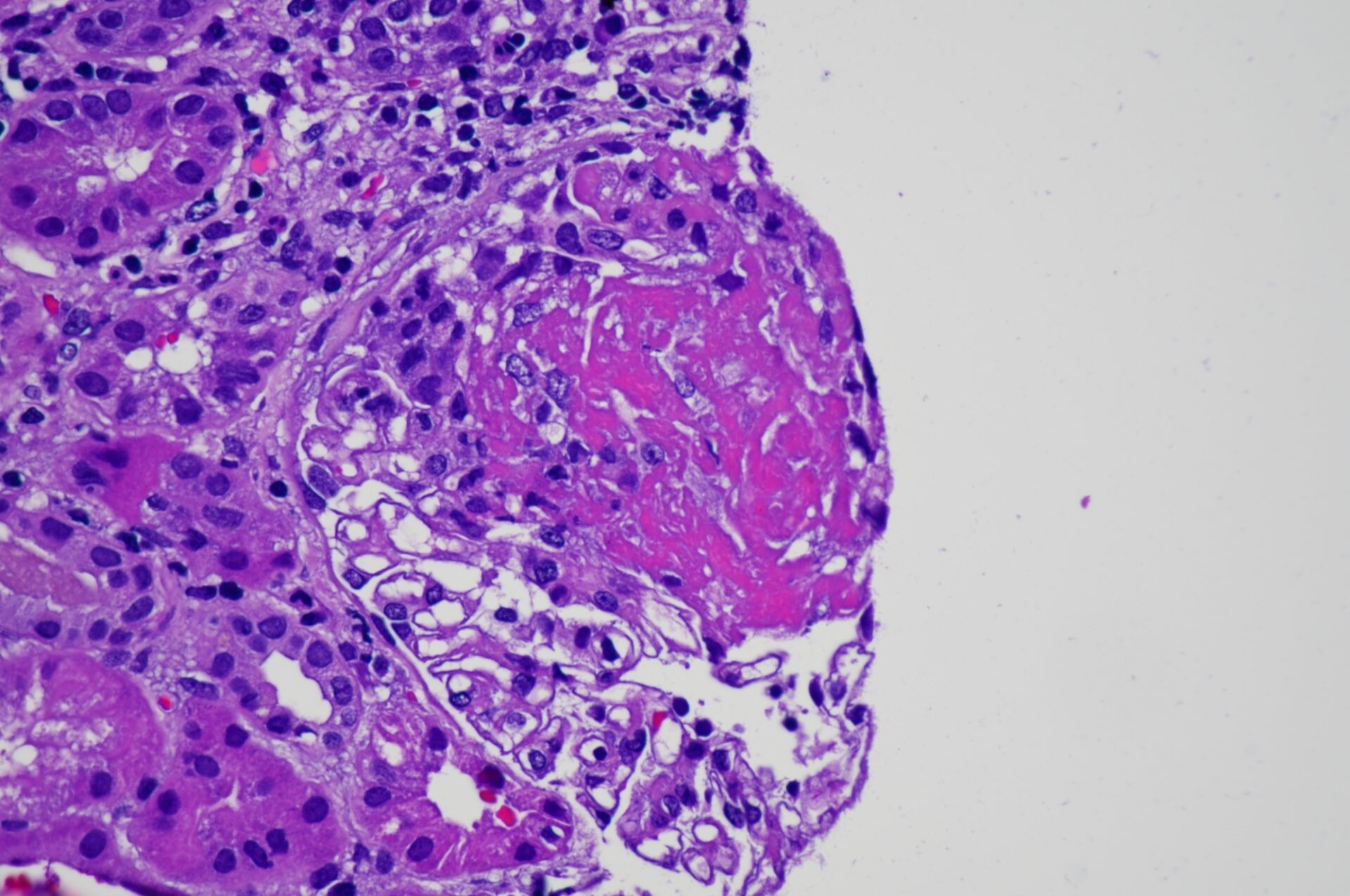
Fibrinoid necrosis in a patient with ANCA-associated glomerulonephritis
From:
Boonyarit Cheunsuchon, Thailand
MD, Associate professor
Acknowledgements
NDT Kidney Art Editor:
Barbara Mara Klinkhammer, Germany
Also interested in submitting an image to NDT? Please contact ndt@era-online.org